 Movie
Movie Controller


+ Open data

- Basic information
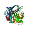
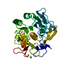
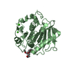
| Entry | Database: PDB / ID: 5gv1 | ||||||
|---|---|---|---|---|---|---|---|
| Title | Crystal structure of ENZbleach xylanase wild type | ||||||
 Components Components | Endo-1,4-beta-xylanase | ||||||
 Keywords Keywords | HYDROLASE / Endo-1 / 4-beta-xylanase / GH11 xylanase | ||||||
| Biological species | termite gut metagenome (others) | ||||||
| Method |  X-RAY DIFFRACTION / MOLECULAR REPLACEMENT / molecular replacement / Resolution: 1.5 Å X-RAY DIFFRACTION / MOLECULAR REPLACEMENT / molecular replacement / Resolution: 1.5 Å | ||||||
| Model details | Crystal structure of ENZbleach xylanase wild type | ||||||
 Authors Authors | Chitnumsub, P. / Jaruwat, A. / Boonyapakorn, K. / Noytanom, K. | ||||||
| Funding support |  Thailand, 1items Thailand, 1items
| ||||||
 Citation Citation | Journal: J. Biotechnol. / Year: 2017 Title: Structure-based protein engineering for thermostable and alkaliphilic enhancement of endo-beta-1,4-xylanase for applications in pulp bleaching Authors: Boonyapakron, K. / Jaruwat, A. / Liwnaree, B. / Nimchua, T. / Champreda, V. / Chitnumsub, P. | ||||||
| History |
|
- Structure visualization
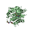
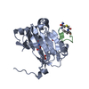
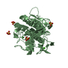
| Structure viewer | Molecule:  MolmilJmol/JSmol MolmilJmol/JSmol |
|---|
- Downloads & links
-Download
| PDBx/mmCIF format | 5gv1.cif.gz | 78.1 KB | Display | PDBx/mmCIF format |
|---|---|---|---|---|
| PDB format | pdb5gv1.ent.gz | 54.9 KB | Display | PDB format |
| PDBx/mmJSON format | 5gv1.json.gz | Tree view | PDBx/mmJSON format | |
| Others |  Other downloads Other downloads |
-Validation report
| Arichive directory | https://data.pdbj.org/pub/pdb/validation_reports/gv/5gv1ftp://data.pdbj.org/pub/pdb/validation_reports/gv/5gv1 | HTTPS FTP |
|---|
-Related structure data
| Related structure data |  5gy8C 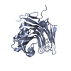 5gy9C 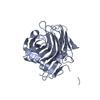 5gyaC 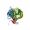 5gybC 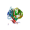 5gycC 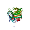 5gyeC 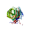 5gyfC  5gygC  5gyhC 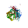 5gyiC 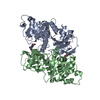 1ig0S S: Starting model for refinement C: citing same article ( |
|---|---|
| Similar structure data |


-Links
 PDBj
PDBj- Assembly
| Deposited unit | 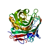
| ||||||||
|---|---|---|---|---|---|---|---|---|---|
| 1 |
| ||||||||
| Unit cell |
|
-Components
| #1: Protein | Mass: 35842.516 Da / Num. of mol.: 1 Source method: isolated from a genetically manipulated source Source: (gene. exp.) termite gut metagenome (others) / Plasmid: pET28a / Production host:  |
|---|---|
| #2: Water | ChemComp-HOH /  Mass: 18.015 Da / Num. of mol.: 416 / Source method: isolated from a natural source / Formula: H2O Mass: 18.015 Da / Num. of mol.: 416 / Source method: isolated from a natural source / Formula: H2O |
| Sequence details | Residues (-35)-0 and 274-286 in the sample sequence are His-tag sequence. Residues 1-273 in the ...Residues (-35)-0 and 274-286 in the sample sequence are His-tag sequence. Residues 1-273 in the sample sequence is the xylanase protein. |
-Experimental details
-Experiment
| Experiment | Method: X-RAY DIFFRACTION / Number of used crystals: 1 |
|---|
- Sample preparation
| Crystal | Density Matthews: 1.9 Å3/Da / Density % sol: 35.38 % / Mosaicity: 0.78 ° |
|---|---|
| Crystal grow | Temperature: 298 K / Method: microbatch / pH: 6.5 / Details: PEG 1500, 0.1 M Bis-Tris |
-Data collection
| Diffraction | Mean temperature: 100 K | ||||||||||||||||||||||||||||||||||||||||||||||||||||||||||||||||||
|---|---|---|---|---|---|---|---|---|---|---|---|---|---|---|---|---|---|---|---|---|---|---|---|---|---|---|---|---|---|---|---|---|---|---|---|---|---|---|---|---|---|---|---|---|---|---|---|---|---|---|---|---|---|---|---|---|---|---|---|---|---|---|---|---|---|---|---|
| Diffraction source | Source: ROTATING ANODE / Type: RIGAKU RUH3R / Wavelength: 1.54 Å | ||||||||||||||||||||||||||||||||||||||||||||||||||||||||||||||||||
| Detector | Type: RIGAKU RAXIS IV++ / Detector: IMAGE PLATE / Date: Jan 14, 2013 / Details: mirrors | ||||||||||||||||||||||||||||||||||||||||||||||||||||||||||||||||||
| Radiation | Monochromator: GRAPHITE / Protocol: SINGLE WAVELENGTH / Monochromatic (M) / Laue (L): M / Scattering type: x-ray | ||||||||||||||||||||||||||||||||||||||||||||||||||||||||||||||||||
| Radiation wavelength | Wavelength: 1.54 Å / Relative weight: 1 | ||||||||||||||||||||||||||||||||||||||||||||||||||||||||||||||||||
| Reflection | Resolution: 1.5→30 Å / Num. obs: 38524 / % possible obs: 91 % / Redundancy: 2.27 % / Biso Wilson estimate: 10.184 Å2 / Rmerge(I) obs: 0.045 / Net I/σ(I): 11.2 | ||||||||||||||||||||||||||||||||||||||||||||||||||||||||||||||||||
| Reflection shell |
|
-Phasing
| Phasing | Method: molecular replacement |
|---|
- Processing
| Software |
| |||||||||||||||||||||||||||||||||||||||||||||
|---|---|---|---|---|---|---|---|---|---|---|---|---|---|---|---|---|---|---|---|---|---|---|---|---|---|---|---|---|---|---|---|---|---|---|---|---|---|---|---|---|---|---|---|---|---|---|
| Refinement | Method to determine structure: MOLECULAR REPLACEMENT Starting model: 1IG0 Resolution: 1.5→29.91 Å / Cor.coef. Fo:Fc: 0.946 / Cor.coef. Fo:Fc free: 0.943 / SU B: 1.234 / SU ML: 0.047 / Cross valid method: THROUGHOUT / σ(F): 0 / ESU R: 0.09 / ESU R Free: 0.081 Details: HYDROGENS HAVE BEEN USED IF PRESENT IN THE INPUT U VALUES : REFINED INDIVIDUALLY
| |||||||||||||||||||||||||||||||||||||||||||||
| Solvent computation | Ion probe radii: 0.8 Å / Shrinkage radii: 0.8 Å / VDW probe radii: 1.4 Å | |||||||||||||||||||||||||||||||||||||||||||||
| Displacement parameters | Biso max: 41.07 Å2 / Biso mean: 9.488 Å2 / Biso min: 4.81 Å2
| |||||||||||||||||||||||||||||||||||||||||||||
| Refinement step | Cycle: final / Resolution: 1.5→29.91 Å
| |||||||||||||||||||||||||||||||||||||||||||||
| Refine LS restraints |
| |||||||||||||||||||||||||||||||||||||||||||||
| LS refinement shell | Resolution: 1.5→1.539 Å / Total num. of bins used: 20
|
