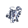
 Movie
Movie Controller


+ Open data

- Basic information
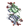
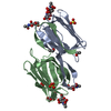
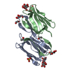
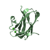
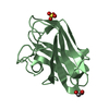
| Entry | Database: PDB / ID: 3ll0 | ||||||
|---|---|---|---|---|---|---|---|
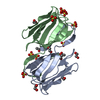
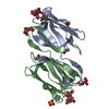
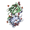
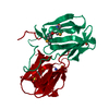
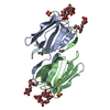
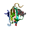
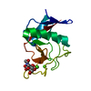
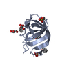
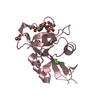
| Title | Monomeric Griffithsin with two Gly-Ser Insertions | ||||||
 Components Components | Griffithsin | ||||||
 Keywords Keywords | SUGAR BINDING PROTEIN / lectin / sugar-binding / anti-HIV / high mannose / Man9 / gp120 / gp41 / jacalin-related / Mannose-binding | ||||||
| Function / homology |  Function and homology information Function and homology informationN-acetylgalactosamine binding / D-glucose binding / D-mannose binding / carbohydrate binding / identical protein binding Similarity search - Function | ||||||
| Biological species |  Griffithsia (eukaryote) Griffithsia (eukaryote) | ||||||
| Method |  X-RAY DIFFRACTION / MOLECULAR REPLACEMENT / Resolution: 1.7 Å X-RAY DIFFRACTION / MOLECULAR REPLACEMENT / Resolution: 1.7 Å | ||||||
 Authors Authors | Moulaei, T. / Wlodawer, A. | ||||||
 Citation Citation | Journal: Structure / Year: 2010 Title: Monomerization of viral entry inhibitor griffithsin elucidates the relationship between multivalent binding to carbohydrates and anti-HIV activity. Authors: Moulaei, T. / Shenoy, S.R. / Giomarelli, B. / Thomas, C. / McMahon, J.B. / Dauter, Z. / O'Keefe, B.R. / Wlodawer, A. | ||||||
| History |
|
- Structure visualization
| Structure viewer | Molecule: MolmilJmol/JSmol |
|---|
- Downloads & links
-Download
| PDBx/mmCIF format | 3ll0.cif.gz | 41.1 KB | Display | PDBx/mmCIF format |
|---|---|---|---|---|
| PDB format | pdb3ll0.ent.gz | 27.9 KB | Display | PDB format |
| PDBx/mmJSON format | 3ll0.json.gz | Tree view | PDBx/mmJSON format | |
| Others |  Other downloads Other downloads |
-Validation report
| Arichive directory | https://data.pdbj.org/pub/pdb/validation_reports/ll/3ll0ftp://data.pdbj.org/pub/pdb/validation_reports/ll/3ll0 | HTTPS FTP |
|---|
-Related structure data
| Related structure data | 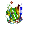 3lkySC 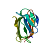 3ll1C 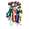 3ll2C S: Starting model for refinement C: citing same article ( |
|---|---|
| Similar structure data |


-Links
 PDBj
PDBj- Assembly
| Deposited unit | 
| ||||||||
|---|---|---|---|---|---|---|---|---|---|
| 1 |
| ||||||||
| Unit cell |
|
-Components
| #1: Protein | Mass: 12989.062 Da / Num. of mol.: 1 / Mutation: Gly-Ser-Gly-Ser inserted after S16 Source method: isolated from a genetically manipulated source Source: (gene. exp.) Griffithsia (eukaryote) / Strain: Q66D336 / Plasmid: pET-15b / Production host:  | ||||
|---|---|---|---|---|---|
| #2: Chemical | ChemComp-GOL /   Mass: 92.094 Da / Num. of mol.: 4 / Source method: obtained synthetically / Formula: C3H8O3 Mass: 92.094 Da / Num. of mol.: 4 / Source method: obtained synthetically / Formula: C3H8O3#3: Chemical | ChemComp-SO4 / |   Mass: 96.063 Da / Num. of mol.: 1 / Source method: obtained synthetically / Formula: SO4 Mass: 96.063 Da / Num. of mol.: 1 / Source method: obtained synthetically / Formula: SO4#4: Water | ChemComp-HOH / |  Mass: 18.015 Da / Num. of mol.: 135 / Source method: isolated from a natural source / Formula: H2O Mass: 18.015 Da / Num. of mol.: 135 / Source method: isolated from a natural source / Formula: H2O |
-Experimental details
-Experiment
| Experiment | Method: X-RAY DIFFRACTION / Number of used crystals: 1 |
|---|
- Sample preparation
| Crystal | Density Matthews: 2.64 Å3/Da / Density % sol: 53.47 % |
|---|---|
| Crystal grow | Temperature: 298 K / Method: vapor diffusion, sitting drop / pH: 4.6 Details: 0.1 M sodium acetate, 0.1 M CdCl, 30% w/v PEG 400, pH 4.6, VAPOR DIFFUSION, SITTING DROP, temperature 298K |
-Data collection
| Diffraction | Mean temperature: 100 K |
|---|---|
| Diffraction source | Source: ROTATING ANODE / Type: RIGAKU MICROMAX-007 HF / Wavelength: 1.5418 Å |
| Detector | Type: MAR scanner 345 mm plate / Detector: IMAGE PLATE / Date: Apr 30, 2009 |
| Radiation | Monochromator: VariMax / Protocol: SINGLE WAVELENGTH / Monochromatic (M) / Laue (L): M / Scattering type: x-ray |
| Radiation wavelength | Wavelength: 1.5418 Å / Relative weight: 1 |
| Reflection | Resolution: 1.7→50 Å / Num. obs: 16094 / % possible obs: 97.9 % / Redundancy: 11.4 % / Rmerge(I) obs: 0.09 / Χ2: 1.003 / Net I/σ(I): 9.5 |
| Reflection shell | Resolution: 1.7→1.76 Å / Redundancy: 5.7 % / Rmerge(I) obs: 0.69 / Mean I/σ(I) obs: 2.1 / Num. unique all: 1478 / Χ2: 0.987 / % possible all: 93.5 |
-Phasing
| Phasing MR | Rfactor: 40.11 / Model details: Phaser MODE: MR_AUTO
|
|---|
- Processing
| Software |
| |||||||||||||||||||||||||||||||||||||||||||||||||||||||||||||||||||||||||||||||||||||
|---|---|---|---|---|---|---|---|---|---|---|---|---|---|---|---|---|---|---|---|---|---|---|---|---|---|---|---|---|---|---|---|---|---|---|---|---|---|---|---|---|---|---|---|---|---|---|---|---|---|---|---|---|---|---|---|---|---|---|---|---|---|---|---|---|---|---|---|---|---|---|---|---|---|---|---|---|---|---|---|---|---|---|---|---|---|---|
| Refinement | Method to determine structure: MOLECULAR REPLACEMENT Starting model: 3LKY Resolution: 1.7→30 Å / Cor.coef. Fo:Fc: 0.958 / Cor.coef. Fo:Fc free: 0.949 / Occupancy max: 1 / Occupancy min: 0.5 / SU B: 3.563 / SU ML: 0.064 / Cross valid method: THROUGHOUT / σ(F): 0 / ESU R: 0.103 / ESU R Free: 0.099 / Stereochemistry target values: MAXIMUM LIKELIHOOD / Details: HYDROGENS HAVE BEEN ADDED IN THE RIDING POSITIONS
| |||||||||||||||||||||||||||||||||||||||||||||||||||||||||||||||||||||||||||||||||||||
| Solvent computation | Ion probe radii: 0.8 Å / Shrinkage radii: 0.8 Å / VDW probe radii: 1.2 Å / Solvent model: MASK | |||||||||||||||||||||||||||||||||||||||||||||||||||||||||||||||||||||||||||||||||||||
| Displacement parameters | Biso max: 57.09 Å2 / Biso mean: 20.174 Å2 / Biso min: 12.14 Å2
| |||||||||||||||||||||||||||||||||||||||||||||||||||||||||||||||||||||||||||||||||||||
| Refinement step | Cycle: LAST / Resolution: 1.7→30 Å
| |||||||||||||||||||||||||||||||||||||||||||||||||||||||||||||||||||||||||||||||||||||
| Refine LS restraints |
| |||||||||||||||||||||||||||||||||||||||||||||||||||||||||||||||||||||||||||||||||||||
| LS refinement shell | Resolution: 1.7→1.743 Å / Total num. of bins used: 20
| |||||||||||||||||||||||||||||||||||||||||||||||||||||||||||||||||||||||||||||||||||||
| Refinement TLS params. | Method: refined / Origin x: 16.806 Å / Origin y: 6.271 Å / Origin z: 18.878 Å
|
