 Movie
Movie Controller

[English] 日本語
 Yorodumi
- PDB-3lis: Crystal Structure of the Restriction-Modification Controller Prot... -
+ Open data

- Basic information
| Entry | Database: PDB / ID: 3lis | ||||||
|---|---|---|---|---|---|---|---|
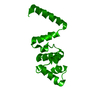
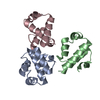
| Title | Crystal Structure of the Restriction-Modification Controller Protein C.Csp231I (Monoclinic form) | ||||||
 Components Components | Csp231I C protein | ||||||
 Keywords Keywords | TRANSCRIPTION / transcriptional regulator / helix-turn-helix / DNA binding protein / restriction modification control | ||||||
| Function / homology |  Function and homology information Function and homology information | ||||||
| Biological species |  Citrobacter sp. RFL231 (bacteria) Citrobacter sp. RFL231 (bacteria) | ||||||
| Method |  X-RAY DIFFRACTION / SYNCHROTRON / MOLECULAR REPLACEMENT / molecular replacement / Resolution: 2 Å X-RAY DIFFRACTION / SYNCHROTRON / MOLECULAR REPLACEMENT / molecular replacement / Resolution: 2 Å | ||||||
 Authors Authors | McGeehan, J.E. / Streeter, S.D. / Thresh, S.J. / Kneale, G.G. | ||||||
 Citation Citation | Journal: J.Mol.Biol. / Year: 2011 Title: Structural Analysis of a Novel Class of R-M Controller Proteins: C.Csp231I from Citrobacter sp. RFL231. Authors: McGeehan, J.E. / Streeter, S.D. / Thresh, S.J. / Taylor, J.E. / Shevtsov, M.B. / Kneale, G.G. | ||||||
| History |
|
- Structure visualization
| Structure viewer | Molecule: MolmilJmol/JSmol |
|---|
- Downloads & links
-Download
| PDBx/mmCIF format | 3lis.cif.gz | 51.6 KB | Display | PDBx/mmCIF format |
|---|---|---|---|---|
| PDB format | pdb3lis.ent.gz | 37.9 KB | Display | PDB format |
| PDBx/mmJSON format | 3lis.json.gz | Tree view | PDBx/mmJSON format | |
| Others |  Other downloads Other downloads |
-Validation report
| Summary document | 3lis_validation.pdf.gz | 423.1 KB | Display | wwPDB validaton report |
|---|---|---|---|---|
| Full document | 3lis_full_validation.pdf.gz | 425.3 KB | Display | |
| Data in XML | 3lis_validation.xml.gz | 10 KB | Display | |
| Data in CIF | 3lis_validation.cif.gz | 13 KB | Display | |
| Arichive directory | https://data.pdbj.org/pub/pdb/validation_reports/li/3lisftp://data.pdbj.org/pub/pdb/validation_reports/li/3lis | HTTPS FTP |
-Related structure data


-Links
 PDBj
PDBj


- Assembly
| Deposited unit | 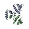
| ||||||||||||||||||
|---|---|---|---|---|---|---|---|---|---|---|---|---|---|---|---|---|---|---|---|
| 1 |
| ||||||||||||||||||
| Unit cell |
| ||||||||||||||||||
| Noncrystallographic symmetry (NCS) | NCS domain:
NCS domain segments: Component-ID: 1 / Ens-ID: 1 / Beg auth comp-ID: MET / Beg label comp-ID: MET / End auth comp-ID: LYS / End label comp-ID: LYS / Refine code: 1 / Auth seq-ID: 1 - 96 / Label seq-ID: 1 - 96
|
-Components
| #1: Protein | Mass: 11380.236 Da / Num. of mol.: 2 Source method: isolated from a genetically manipulated source Source: (gene. exp.) Citrobacter sp. RFL231 (bacteria) / Gene: csp231IC / Plasmid: pET11a / Production host: #2: Water | ChemComp-HOH / |  Mass: 18.015 Da / Num. of mol.: 88 / Source method: isolated from a natural source / Formula: H2O Mass: 18.015 Da / Num. of mol.: 88 / Source method: isolated from a natural source / Formula: H2O |
|---|
-Experimental details
-Experiment
| Experiment | Method: X-RAY DIFFRACTION / Number of used crystals: 1 |
|---|
- Sample preparation
| Crystal | Density Matthews: 2 Å3/Da / Density % sol: 38.59 % |
|---|---|
| Crystal grow | Temperature: 289 K / Method: vapor diffusion, hanging drop / pH: 7 Details: 0.1M malate-MES-Tris (MMT), 20% PEG 1500, pH 7.0, VAPOR DIFFUSION, HANGING DROP, temperature 289K |
-Data collection
| Diffraction | Mean temperature: 100 K |
|---|---|
| Diffraction source | Source: SYNCHROTRON / Site: Diamond  / Beamline: I02 / Wavelength: 0.9795 Å / Beamline: I02 / Wavelength: 0.9795 Å |
| Detector | Type: ADSC QUANTUM 315 / Detector: CCD / Date: Apr 7, 2009 |
| Radiation | Monochromator: Double crystal / Protocol: SINGLE WAVELENGTH / Monochromatic (M) / Laue (L): M / Scattering type: x-ray |
| Radiation wavelength | Wavelength: 0.9795 Å / Relative weight: 1 |
| Reflection | Resolution: 2→50 Å / Num. obs: 12439 / % possible obs: 99.2 % / Observed criterion σ(F): 0 / Observed criterion σ(I): 0 / Redundancy: 3.3 % / Rsym value: 0.057 / Net I/σ(I): 11.7 |
| Reflection shell | Resolution: 2→2.11 Å / Redundancy: 3.4 % / Rmerge(I) obs: 0.276 / Mean I/σ(I) obs: 4.1 / Num. unique all: 1821 / % possible all: 99.7 |
-Phasing
| Phasing | Method: molecular replacement | |||||||||
|---|---|---|---|---|---|---|---|---|---|---|
| Phasing MR | Rfactor: 33.96 / Model details: Phaser MODE: MR_AUTO
|
- Processing
| Software |
| |||||||||||||||||||||||||||||||||||||||||||||||||||||||||||||||||
|---|---|---|---|---|---|---|---|---|---|---|---|---|---|---|---|---|---|---|---|---|---|---|---|---|---|---|---|---|---|---|---|---|---|---|---|---|---|---|---|---|---|---|---|---|---|---|---|---|---|---|---|---|---|---|---|---|---|---|---|---|---|---|---|---|---|---|
| Refinement | Method to determine structure: MOLECULAR REPLACEMENT / Resolution: 2→31.5 Å / Cor.coef. Fo:Fc: 0.942 / Cor.coef. Fo:Fc free: 0.921 / WRfactor Rfree: 0.255 / WRfactor Rwork: 0.213 / Occupancy max: 1 / Occupancy min: 1 / FOM work R set: 0.843 / SU R Cruickshank DPI: 0.24 / SU Rfree: 0.189 / Cross valid method: THROUGHOUT / σ(F): 0 / ESU R: 0.24 / ESU R Free: 0.189 / Stereochemistry target values: MAXIMUM LIKELIHOOD Details: HYDROGENS HAVE BEEN ADDED IN THE RIDING POSITIONS. U VALUES REFINED INDIVIDUALLY
| |||||||||||||||||||||||||||||||||||||||||||||||||||||||||||||||||
| Solvent computation | Ion probe radii: 0.8 Å / Shrinkage radii: 0.8 Å / VDW probe radii: 1.4 Å / Solvent model: MASK | |||||||||||||||||||||||||||||||||||||||||||||||||||||||||||||||||
| Displacement parameters | Biso max: 76.97 Å2 / Biso mean: 33.417 Å2 / Biso min: 14.21 Å2
| |||||||||||||||||||||||||||||||||||||||||||||||||||||||||||||||||
| Refinement step | Cycle: LAST / Resolution: 2→31.5 Å
| |||||||||||||||||||||||||||||||||||||||||||||||||||||||||||||||||
| Refine LS restraints |
| |||||||||||||||||||||||||||||||||||||||||||||||||||||||||||||||||
| Refine LS restraints NCS | Dom-ID: 1 / Auth asym-ID: A / Ens-ID: 1 / Number: 784 / Refine-ID: X-RAY DIFFRACTION
| |||||||||||||||||||||||||||||||||||||||||||||||||||||||||||||||||
| LS refinement shell | Resolution: 2→2.052 Å / Total num. of bins used: 20
|
