 Movie
Movie Controller

[English] 日本語
 Yorodumi
- PDB-3lhb: THE 2.7 ANGSTROM CRYSTAL STRUCTURE OF DEOXYGENATED HEMOGLOBIN FRO... -
+ Open data

- Basic information
| Entry | Database: PDB / ID: 3lhb | ||||||
|---|---|---|---|---|---|---|---|
| Title | THE 2.7 ANGSTROM CRYSTAL STRUCTURE OF DEOXYGENATED HEMOGLOBIN FROM THE SEA LAMPREY (PETROMYZON MARINUS) | ||||||
 Components Components | PROTEIN (HEMOGLOBIN) | ||||||
 Keywords Keywords | METAL BINDING PROTEIN / HEMOGLOBIN / OXYGEN TRANSPORT / BOHR EFFECT | ||||||
| Function / homology |  Function and homology information Function and homology informationoxygen carrier activity / oxygen binding / oxidoreductase activity / iron ion binding / heme binding Similarity search - Function | ||||||
| Biological species |  | ||||||
| Method |  X-RAY DIFFRACTION / MOLECULAR REPLACEMENT / Resolution: 2.7 Å X-RAY DIFFRACTION / MOLECULAR REPLACEMENT / Resolution: 2.7 Å | ||||||
 Authors Authors | Heaslet, H.A. / Royer Jr., W.E. | ||||||
 Citation Citation | Journal: Structure Fold.Des. / Year: 1999 Title: The 2.7 A crystal structure of deoxygenated hemoglobin from the sea lamprey (Petromyzon marinus): structural basis for a lowered oxygen affinity and Bohr effect. Authors: Heaslet, H.A. / Royer Jr., W.E. #1: Journal: J.Mol.Biol. / Year: 1985Title: Refinement of a Molcular Model for Lamprey Hemoglobin from Petromyzon Marinus Authors: Honzatko, R.B. / Hendrickson, W.A. / Love, W.E. #2: Journal: Nature New Biol. / Year: 1971Title: Structure of Lamprey Hemoglobin Authors: Hendrickson, W.A. / Love, W.E. | ||||||
| History |
|
- Structure visualization
| Structure viewer | Molecule: MolmilJmol/JSmol |
|---|
- Downloads & links
-Download
| PDBx/mmCIF format | 3lhb.cif.gz | 319.1 KB | Display | PDBx/mmCIF format |
|---|---|---|---|---|
| PDB format | pdb3lhb.ent.gz | 272.1 KB | Display | PDB format |
| PDBx/mmJSON format | 3lhb.json.gz | Tree view | PDBx/mmJSON format | |
| Others |  Other downloads Other downloads |
-Validation report
| Arichive directory | https://data.pdbj.org/pub/pdb/validation_reports/lh/3lhbftp://data.pdbj.org/pub/pdb/validation_reports/lh/3lhb | HTTPS FTP |
|---|
-Related structure data
| Related structure data | 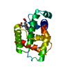 2lhbS S: Starting model for refinement |
|---|---|
| Similar structure data |


-Links
 PDBj
PDBj


- Assembly
| Deposited unit | 
| ||||||||||||||||||||||||||||||||||||||||||||||||
|---|---|---|---|---|---|---|---|---|---|---|---|---|---|---|---|---|---|---|---|---|---|---|---|---|---|---|---|---|---|---|---|---|---|---|---|---|---|---|---|---|---|---|---|---|---|---|---|---|---|
| 1 | 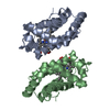
| ||||||||||||||||||||||||||||||||||||||||||||||||
| 2 | 
| ||||||||||||||||||||||||||||||||||||||||||||||||
| 3 | 
| ||||||||||||||||||||||||||||||||||||||||||||||||
| 4 | 
| ||||||||||||||||||||||||||||||||||||||||||||||||
| 5 | 
| ||||||||||||||||||||||||||||||||||||||||||||||||
| 6 | 
| ||||||||||||||||||||||||||||||||||||||||||||||||
| Unit cell |
| ||||||||||||||||||||||||||||||||||||||||||||||||
| Noncrystallographic symmetry (NCS) | NCS oper:
| ||||||||||||||||||||||||||||||||||||||||||||||||
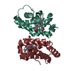
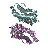
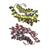
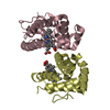
| Details | THE TWELVE PROTOMERS IN THE ASYMMETRIC UNIT FORM SIX ESSENTIALLY IDENTICAL DIMERS. |
-Components
| #1: Protein | Mass: 16291.678 Da / Num. of mol.: 12 / Source method: isolated from a natural source Details: COMPONENT FIVE(5) OF SIX(6) HEMOGLOBINS ISOLATED FROM P.MARINUS. Source: (natural) #2: Chemical | ChemComp-HEM /   Mass: 616.487 Da / Num. of mol.: 12 / Source method: obtained synthetically / Formula: C34H32FeN4O4 Mass: 616.487 Da / Num. of mol.: 12 / Source method: obtained synthetically / Formula: C34H32FeN4O4 |
|---|
-Experimental details
-Experiment
| Experiment | Method: X-RAY DIFFRACTION / Number of used crystals: 1 |
|---|
- Sample preparation
| Crystal | Density Matthews: 2.46 Å3/Da / Density % sol: 47 % | |||||||||||||||
|---|---|---|---|---|---|---|---|---|---|---|---|---|---|---|---|---|
| Crystal grow | pH: 6.8 / Details: pH 6.8 | |||||||||||||||
| Crystal | *PLUS | |||||||||||||||
| Crystal grow | *PLUS Method: batch method | |||||||||||||||
| Components of the solutions | *PLUS
|
-Data collection
| Diffraction | Mean temperature: 293.15 K |
|---|---|
| Diffraction source | Source: ROTATING ANODE / Type: RIGAKU RU200 / Wavelength: 1.54 |
| Detector | Type: RIGAKU RAXIS IIC / Detector: IMAGE PLATE / Date: Dec 1, 1997 |
| Radiation | Monochromator: GRAPHITE / Protocol: SINGLE WAVELENGTH / Monochromatic (M) / Laue (L): M / Scattering type: x-ray |
| Radiation wavelength | Wavelength: 1.54 Å / Relative weight: 1 |
| Reflection | Resolution: 2.7→10 Å / Num. obs: 49184 / % possible obs: 94.7 % / Observed criterion σ(I): 2 / Redundancy: 3 % / Rsym value: 0.076 / Net I/σ(I): 12 |
| Reflection shell | Resolution: 2.7→2.8 Å / Redundancy: 3 % / Mean I/σ(I) obs: 2.7 / Rsym value: 0.422 / % possible all: 87.2 |
| Reflection | *PLUS Num. measured all: 147754 / Rmerge(I) obs: 0.076 |
| Reflection shell | *PLUS % possible obs: 87.2 % |
- Processing
| Software |
| ||||||||||||||||||||||||||||||||||||||||||||||||||||||||||||
|---|---|---|---|---|---|---|---|---|---|---|---|---|---|---|---|---|---|---|---|---|---|---|---|---|---|---|---|---|---|---|---|---|---|---|---|---|---|---|---|---|---|---|---|---|---|---|---|---|---|---|---|---|---|---|---|---|---|---|---|---|---|
| Refinement | Method to determine structure: MOLECULAR REPLACEMENT Starting model: 2LHB Resolution: 2.7→10 Å / Isotropic thermal model: GROUP THERMAL FACTORS / Cross valid method: THROUGHOUT / σ(F): 2
| ||||||||||||||||||||||||||||||||||||||||||||||||||||||||||||
| Displacement parameters | Biso mean: 44.5 Å2 | ||||||||||||||||||||||||||||||||||||||||||||||||||||||||||||
| Refinement step | Cycle: LAST / Resolution: 2.7→10 Å
| ||||||||||||||||||||||||||||||||||||||||||||||||||||||||||||
| Refine LS restraints |
| ||||||||||||||||||||||||||||||||||||||||||||||||||||||||||||
| Refine LS restraints NCS | NCS model details: RESTRAINTS / Rms dev position: 1.5 Å / Weight position: 300 | ||||||||||||||||||||||||||||||||||||||||||||||||||||||||||||
| LS refinement shell | Resolution: 2.7→2.8 Å / Total num. of bins used: 8
| ||||||||||||||||||||||||||||||||||||||||||||||||||||||||||||
| Xplor file |
| ||||||||||||||||||||||||||||||||||||||||||||||||||||||||||||
| Software | *PLUS Name: X-PLOR / Version: 3.851 / Classification: refinement | ||||||||||||||||||||||||||||||||||||||||||||||||||||||||||||
| Refinement | *PLUS Highest resolution: 2.7 Å / Lowest resolution: 10 Å / σ(F): 2 / % reflection Rfree: 11.4 % | ||||||||||||||||||||||||||||||||||||||||||||||||||||||||||||
| Solvent computation | *PLUS | ||||||||||||||||||||||||||||||||||||||||||||||||||||||||||||
| Displacement parameters | *PLUS Biso mean: 44.5 Å2 | ||||||||||||||||||||||||||||||||||||||||||||||||||||||||||||
| Refine LS restraints | *PLUS
| ||||||||||||||||||||||||||||||||||||||||||||||||||||||||||||
| LS refinement shell | *PLUS Highest resolution: 2.7 Å / Lowest resolution: 2.8 Å / Rfactor Rfree: 0.329 / % reflection Rfree: 10.9 % / Rfactor Rwork: 0.287 |
