 ムービー
ムービー コントローラー


+ データを開く

- 基本情報
基本情報
| 登録情報 | データベース: PDB / ID: 1f5o | ||||||
|---|---|---|---|---|---|---|---|
| タイトル | 2.9 ANGSTROM CRYSTAL STRUCTURE OF DEOXYGENATED LAMPREY HEMOGLOBIN V IN THE SPACE GROUP P2(1)2(1)2(1) | ||||||
 要素 要素 | HEMOGLOBIN V | ||||||
 キーワード キーワード | OXYGEN STORAGE/TRANSPORT / Hemoglobin / Heme / Lamprey / OXYGEN STORAGE-TRANSPORT COMPLEX | ||||||
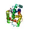
| 機能・相同性 |  機能・相同性情報 機能・相同性情報oxygen carrier activity / oxygen binding / oxidoreductase activity / iron ion binding / heme binding 類似検索 - 分子機能 | ||||||
| 生物種 |  | ||||||
| 手法 |  X線回折 / 解像度: 2.9 Å X線回折 / 解像度: 2.9 Å | ||||||
 データ登録者 データ登録者 | Heaslet, H.A. / Royer Jr., W.E. | ||||||
 引用 引用 | ジャーナル: J.Biol.Chem. / 年: 2001 タイトル: Crystalline ligand transitions in lamprey hemoglobin. Structural evidence for the regulation of oxygen affinity. 著者: Heaslet, H.A. / Royer Jr., W.E. #1: ジャーナル: Structure / 年: 1999タイトル: The 2.7 Angstrom Crystal Structure of Deoxygentated Hemoglobin from the Sea Lamprey (Petromyzon Marinus): Structural Basis for a Lowered Oxygen Affinity and Bohr Effect. 著者: Heaslet, H.A. / Royer Jr., W.E. | ||||||
| 履歴 |
|
- 構造の表示
構造の表示
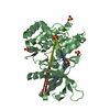
| 構造ビューア | 分子: MolmilJmol/JSmol |
|---|
- ダウンロードとリンク
-ダウンロード
| PDBx/mmCIF形式 | 1f5o.cif.gz | 166.3 KB | 表示 | PDBx/mmCIF形式 |
|---|---|---|---|---|
| PDB形式 | pdb1f5o.ent.gz | 138.5 KB | 表示 | PDB形式 |
| PDBx/mmJSON形式 | 1f5o.json.gz | ツリー表示 | PDBx/mmJSON形式 | |
| その他 |  その他のダウンロード その他のダウンロード |
-検証レポート
| アーカイブディレクトリ | https://data.pdbj.org/pub/pdb/validation_reports/f5/1f5oftp://data.pdbj.org/pub/pdb/validation_reports/f5/1f5o | HTTPS FTP |
|---|
-関連構造データ


-リンク
 PDBj
PDBj


- 集合体
集合体
| 登録構造単位 | 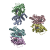
| ||||||||
|---|---|---|---|---|---|---|---|---|---|
| 1 | 
| ||||||||
| 2 | 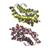
| ||||||||
| 3 | 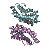
| ||||||||
| 単位格子 |
| ||||||||
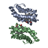
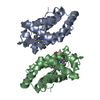
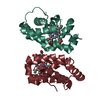
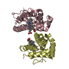
| 詳細 | The physiologically relevant assembly is a homodimer in which the subunits are related by a twofold axis of symmetry. / A hexameric assembly is observed upon application of crystallographic symmetry operators. The subunits in the hexamer are arranged as one turn of an approximately three-fold screw. |
-要素
| #1: タンパク質 | 分子量: 16289.707 Da / 分子数: 6 / 由来タイプ: 天然 / 由来: (天然) #2: 化合物 | ChemComp-HEM /   分子量: 616.487 Da / 分子数: 6 / 由来タイプ: 合成 / 式: C34H32FeN4O4 分子量: 616.487 Da / 分子数: 6 / 由来タイプ: 合成 / 式: C34H32FeN4O4#3: 水 | ChemComp-HOH / |  分子量: 18.015 Da / 分子数: 12 / 由来タイプ: 天然 / 式: H2O 分子量: 18.015 Da / 分子数: 12 / 由来タイプ: 天然 / 式: H2O |
|---|
-実験情報
-実験
| 実験 | 手法: X線回折 / 使用した結晶の数: 1 |
|---|
- 試料調製
試料調製
| 結晶 | マシュー密度: 2.96 Å3/Da / 溶媒含有率: 58.51 % | |||||||||||||||
|---|---|---|---|---|---|---|---|---|---|---|---|---|---|---|---|---|
| 結晶化 | 温度: 296 K / 手法: small tubes / pH: 6.8 詳細: 25% PEG 4K, 170mM phosphate buffer, pH 6.8, SMALL TUBES, temperature 296K | |||||||||||||||
| 結晶化 | *PLUS 手法: batch method | |||||||||||||||
| 溶液の組成 | *PLUS
|
-データ収集
| 回折 | 平均測定温度: 296 K |
|---|---|
| 放射光源 | 由来: 回転陽極 / タイプ: RIGAKU RU200 / 波長: 1.5418 |
| 検出器 | タイプ: RIGAKU RAXIS IV / 検出器: IMAGE PLATE / 日付: 1999年5月17日 |
| 放射 | プロトコル: SINGLE WAVELENGTH / 単色(M)・ラウエ(L): M / 散乱光タイプ: x-ray |
| 放射波長 | 波長: 1.5418 Å / 相対比: 1 |
| 反射 | 解像度: 2.9→40 Å / Num. all: 138470 / Num. obs: 25042 / % possible obs: 95.5 % / Observed criterion σ(F): 0 / Observed criterion σ(I): 2 / 冗長度: 5.5 % / Biso Wilson estimate: 34.2 Å2 / Rmerge(I) obs: 0.081 / Net I/σ(I): 13.8 |
| 反射 シェル | 解像度: 2.9→3 Å / 冗長度: 10.1 % / Rmerge(I) obs: 0.369 / Num. unique all: 2472 / % possible all: 95.1 |
| 反射 | *PLUS Num. measured all: 138470 |
| 反射 シェル | *PLUS % possible obs: 95.4 % / Mean I/σ(I) obs: 5.6 |
- 解析
解析
| ソフトウェア |
| |||||||||||||||||||||||||
|---|---|---|---|---|---|---|---|---|---|---|---|---|---|---|---|---|---|---|---|---|---|---|---|---|---|---|
| 精密化 | 解像度: 2.9→8 Å / σ(F): 0 / σ(I): 2 / 立体化学のターゲット値: Engh & Huber 詳細: Simulated annealing, Powell minimization and group B-factor refinements using tight non-crystallographic symmetry restraints (wa=300). ALTERNATE CRYSTAL FORM FOR DEOXY LHBV WITH SIX MONOMERS ...詳細: Simulated annealing, Powell minimization and group B-factor refinements using tight non-crystallographic symmetry restraints (wa=300). ALTERNATE CRYSTAL FORM FOR DEOXY LHBV WITH SIX MONOMERS IN THE ASYMMETRIC UNIT.
| |||||||||||||||||||||||||
| 精密化ステップ | サイクル: LAST / 解像度: 2.9→8 Å
| |||||||||||||||||||||||||
| 拘束条件 |
| |||||||||||||||||||||||||
| ソフトウェア | *PLUS 名称: CNS / バージョン: 0.9 / 分類: refinement | |||||||||||||||||||||||||
| 精密化 | *PLUS 最高解像度: 2.9 Å / 最低解像度: 8 Å / σ(F): 0 / Rfactor obs: 0.192 | |||||||||||||||||||||||||
| 溶媒の処理 | *PLUS | |||||||||||||||||||||||||
| 原子変位パラメータ | *PLUS |
