 Movie
Movie Controller

[English] 日本語
 Yorodumi
- PDB-3l89: Human Adenovirus type 21 knob in complex with domains SCR1 and SC... -
+ Open data

- Basic information
| Entry | Database: PDB / ID: 3l89 | ||||||
|---|---|---|---|---|---|---|---|
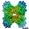
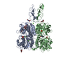
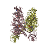
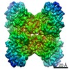
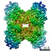
| Title | Human Adenovirus type 21 knob in complex with domains SCR1 and SCR2 of CD46 (membrane cofactor protein, MCP) | ||||||
 Components Components |
| ||||||
 Keywords Keywords | VIRAL PROTEIN/PROTEIN BINDING / Adenovirus / Fiber Knob / Viral Protein / Membrane cofactor protein / MCP / virus receptor complex / SCR / Short consensus repeat / CCP / complement control protein / Complement pathway / Glycoprotein / Host-virus interaction / Immune response / Innate immunity / Sushi2 / VIRAL PROTEIN-PROTEIN BINDING complex | ||||||
| Function / homology |  Function and homology information Function and homology information: / inner acrosomal membrane / negative regulation of complement activation, classical pathway / positive regulation of transforming growth factor beta production / T cell mediated immunity / regulation of Notch signaling pathway / positive regulation of memory T cell differentiation / adhesion receptor-mediated virion attachment to host cell / positive regulation of regulatory T cell differentiation / positive regulation of interleukin-10 production ...: / inner acrosomal membrane / negative regulation of complement activation, classical pathway / positive regulation of transforming growth factor beta production / T cell mediated immunity / regulation of Notch signaling pathway / positive regulation of memory T cell differentiation / adhesion receptor-mediated virion attachment to host cell / positive regulation of regulatory T cell differentiation / positive regulation of interleukin-10 production / complement activation, classical pathway / single fertilization / positive regulation of T cell proliferation / Regulation of Complement cascade / viral capsid / virus receptor activity / signaling receptor activity / adaptive immune response / cell adhesion / cadherin binding / negative regulation of gene expression / innate immune response / focal adhesion / symbiont entry into host cell / positive regulation of gene expression / host cell nucleus / cell surface / : / extracellular exosome / plasma membrane Similarity search - Function | ||||||
| Biological species |  Human adenovirus 21 Human adenovirus 21 Homo sapiens (human) Homo sapiens (human) | ||||||
| Method |  X-RAY DIFFRACTION / SYNCHROTRON / MOLECULAR REPLACEMENT / Resolution: 3.5 Å X-RAY DIFFRACTION / SYNCHROTRON / MOLECULAR REPLACEMENT / Resolution: 3.5 Å | ||||||
 Authors Authors | Cupelli, K. / Stehle, T. | ||||||
 Citation Citation | Journal: J.Virol. / Year: 2010 Title: Structure of adenovirus type 21 knob in complex with CD46 reveals key differences in receptor contacts among species B adenoviruses. Authors: Cupelli, K. / Muller, S. / Persson, B.D. / Jost, M. / Arnberg, N. / Stehle, T. | ||||||
| History |
|
- Structure visualization
| Structure viewer | Molecule: MolmilJmol/JSmol |
|---|
- Downloads & links
-Download
| PDBx/mmCIF format | 3l89.cif.gz | 700.5 KB | Display | PDBx/mmCIF format |
|---|---|---|---|---|
| PDB format | pdb3l89.ent.gz | 580.9 KB | Display | PDB format |
| PDBx/mmJSON format | 3l89.json.gz | Tree view | PDBx/mmJSON format | |
| Others |  Other downloads Other downloads |
-Validation report
| Arichive directory | https://data.pdbj.org/pub/pdb/validation_reports/l8/3l89ftp://data.pdbj.org/pub/pdb/validation_reports/l8/3l89 | HTTPS FTP |
|---|
-Related structure data
| Related structure data |  3l88C  2o39S S: Starting model for refinement C: citing same article ( |
|---|---|
| Similar structure data |


-Links
 PDBj
PDBj

- Assembly
| Deposited unit | 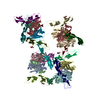
| ||||||||||||||||||||||||||||||||||||||||||||||||||||||||||||||||||||||||||||||||||||||||||||||||||||||||||||||||||||||
|---|---|---|---|---|---|---|---|---|---|---|---|---|---|---|---|---|---|---|---|---|---|---|---|---|---|---|---|---|---|---|---|---|---|---|---|---|---|---|---|---|---|---|---|---|---|---|---|---|---|---|---|---|---|---|---|---|---|---|---|---|---|---|---|---|---|---|---|---|---|---|---|---|---|---|---|---|---|---|---|---|---|---|---|---|---|---|---|---|---|---|---|---|---|---|---|---|---|---|---|---|---|---|---|---|---|---|---|---|---|---|---|---|---|---|---|---|---|---|---|
| 1 | 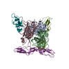
| ||||||||||||||||||||||||||||||||||||||||||||||||||||||||||||||||||||||||||||||||||||||||||||||||||||||||||||||||||||||
| 2 | 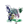
| ||||||||||||||||||||||||||||||||||||||||||||||||||||||||||||||||||||||||||||||||||||||||||||||||||||||||||||||||||||||
| 3 | 
| ||||||||||||||||||||||||||||||||||||||||||||||||||||||||||||||||||||||||||||||||||||||||||||||||||||||||||||||||||||||
| 4 | 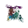
| ||||||||||||||||||||||||||||||||||||||||||||||||||||||||||||||||||||||||||||||||||||||||||||||||||||||||||||||||||||||
| Unit cell |
| ||||||||||||||||||||||||||||||||||||||||||||||||||||||||||||||||||||||||||||||||||||||||||||||||||||||||||||||||||||||
| Noncrystallographic symmetry (NCS) | NCS domain:
NCS domain segments:
|