[日本語] English
- PDB-3kt2: Crystal Structure of N88D mutant HIV-1 Protease -

+
データを開く


IDまたはキーワード:

読み込み中...

-
基本情報

登録情報
データベース: PDB / ID: 3kt2
タイトルCrystal Structure of N88D mutant HIV-1 Protease
要素Protease
キーワードHYDROLASE / Drug Resistant Mutation / N88D / Nelfinavir / HIV-1 protease / Protease
機能・相同性
機能・相同性情報


integrase activity / Integration of viral DNA into host genomic DNA / Autointegration results in viral DNA circles / Minus-strand DNA synthesis / Plus-strand DNA synthesis / 2-LTR circle formation / Uncoating of the HIV Virion / Vpr-mediated nuclear import of PICs / Early Phase of HIV Life Cycle / Integration of provirus ...integrase activity / Integration of viral DNA into host genomic DNA / Autointegration results in viral DNA circles / Minus-strand DNA synthesis / Plus-strand DNA synthesis / 2-LTR circle formation / Uncoating of the HIV Virion / Vpr-mediated nuclear import of PICs / Early Phase of HIV Life Cycle / Integration of provirus / APOBEC3G mediated resistance to HIV-1 infection / Binding and entry of HIV virion / viral life cycle / Assembly Of The HIV Virion / HIV-1 retropepsin / retroviral ribonuclease H / exoribonuclease H / Budding and maturation of HIV virion / exoribonuclease H activity / protein processing / host multivesicular body / viral genome integration into host DNA / RNA-directed DNA polymerase / establishment of integrated proviral latency / viral penetration into host nucleus / RNA stem-loop binding / RNA-directed DNA polymerase activity / host cell / RNA-DNA hybrid ribonuclease activity / 転移酵素; リンを含む基を移すもの; 核酸を移すもの / peptidase activity / symbiont-mediated suppression of host gene expression / viral nucleocapsid / DNA recombination / DNA-directed DNA polymerase / 加水分解酵素; エステル加水分解酵素 / aspartic-type endopeptidase activity / DNA-directed DNA polymerase activity / symbiont entry into host cell / lipid binding / host cell nucleus / host cell plasma membrane / virion membrane / structural molecule activity / DNA binding / zinc ion binding / identical protein binding / membrane
類似検索 - 分子機能
Reverse transcriptase connection / Reverse transcriptase connection domain / Reverse transcriptase thumb / Reverse transcriptase thumb domain / Integrase Zinc binding domain / Zinc finger integrase-type profile. / Integrase-like, N-terminal / Integrase DNA binding domain / Integrase, C-terminal domain superfamily, retroviral / Integrase, N-terminal zinc-binding domain ...Reverse transcriptase connection / Reverse transcriptase connection domain / Reverse transcriptase thumb / Reverse transcriptase thumb domain / Integrase Zinc binding domain / Zinc finger integrase-type profile. / Integrase-like, N-terminal / Integrase DNA binding domain / Integrase, C-terminal domain superfamily, retroviral / Integrase, N-terminal zinc-binding domain / Integrase, C-terminal, retroviral / Integrase DNA binding domain profile. / Immunodeficiency lentiviral matrix, N-terminal / gag gene protein p17 (matrix protein) / RNase H / Integrase core domain / Integrase, catalytic core / Integrase catalytic domain profile. / Retroviral nucleocapsid Gag protein p24, C-terminal domain / Gag protein p24 C-terminal domain / Retropepsin-like catalytic domain / Matrix protein, lentiviral and alpha-retroviral, N-terminal / Ribonuclease H domain / RNase H type-1 domain profile. / Reverse transcriptase (RNA-dependent DNA polymerase) / Reverse transcriptase domain / Reverse transcriptase (RT) catalytic domain profile. / Retropepsins / Retroviral aspartyl protease / Aspartyl protease, retroviral-type family profile. / Peptidase A2A, retrovirus, catalytic / Retrovirus capsid, C-terminal / Retroviral matrix protein / Retrovirus capsid, N-terminal / zinc finger / Zinc knuckle / Zinc finger, CCHC-type superfamily / Cathepsin D, subunit A; domain 1 / Acid Proteases / Zinc finger, CCHC-type / Zinc finger CCHC-type profile. / Aspartic peptidase, active site / Eukaryotic and viral aspartyl proteases active site. / Ribonuclease H superfamily / Aspartic peptidase domain superfamily / Ribonuclease H-like superfamily / Reverse transcriptase/Diguanylate cyclase domain / DNA/RNA polymerase superfamily / Beta Barrel / Mainly Beta
類似検索 - ドメイン・相同性
Gag-Pol polyprotein
類似検索 - 構成要素
生物種Human immunodeficiency virus type 1 (ヒト免疫不全ウイルス)
手法X線回折 / シンクロトロン / フーリエ合成 / 解像度: 1.651 Å
データ登録者Bihani, S.C. / Das, A. / Prashar, V. / Ferrer, J.L. / Hosur, M.V.
引用ジャーナル: Biochem.Biophys.Res.Commun. / : 2009
タイトル: Resistance mechanism revealed by crystal structures of unliganded nelfinavir-resistant HIV-1 protease non-active site mutants N88D and N88S.
著者: Bihani, S.C. / Das, A. / Prashar, V. / Ferrer, J.L. / Hosur, M.V.
履歴
登録2009年11月24日登録サイト: RCSB / 処理サイト: PDBJ
改定 1.02010年2月16日Provider: repository / タイプ: Initial release
改定 1.12011年7月13日Group: Version format compliance
改定 1.22017年8月23日Group: Source and taxonomy / カテゴリ: entity_src_gen
改定 1.32021年11月10日Group: Database references / カテゴリ: database_2 / struct_ref_seq_dif
Item: _database_2.pdbx_DOI / _database_2.pdbx_database_accession / _struct_ref_seq_dif.details
改定 1.42024年5月29日Group: Data collection / カテゴリ: chem_comp_atom / chem_comp_bond

-
構造の表示

構造ビューア分子:
MolmilJmol/JSmol

ダウンロードとリンク

-
集合体

登録構造単位
A: Protease


分子量 (理論値)分子数
合計 (水以外)21,9651
ポリマ-21,9651
非ポリマー00
5,206289
1


  • 登録構造と同一
  • 登録者・ソフトウェアが定義した集合体
タイプ名称対称操作
identity operation1_555x,y,z1
手法PISA
単位格子
Length a, b, c (Å)62.050, 62.050, 83.700
Angle α, β, γ (deg.)90.000, 90.000, 120.000
Int Tables number169
Space group name H-MP61

-
要素

#1: タンパク質 Protease


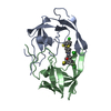
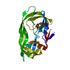
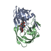
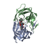
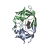
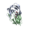
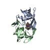
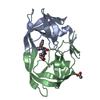
分子量: 21964.848 Da / 分子数: 1 / 変異: N88D, C95M, N1088D, C1095M / 由来タイプ: 組換発現
詳細: The fusion protein of two same subunits (UNP residues 489-587) and linker peptide GGSSG. Two subunits are linked through the linker.
由来: (組換発現) Human immunodeficiency virus type 1 (ヒト免疫不全ウイルス)
遺伝子: gag-pol / プラスミド: pET11A / 発現宿主: Escherichia coli (大腸菌) / 株 (発現宿主): BL21(DE3) / 参照: UniProt: P04585, HIV-1 retropepsin
#2: 水 ChemComp-HOH / water


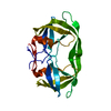
分子量: 18.015 Da / 分子数: 289 / 由来タイプ: 天然 / : H2O
配列の詳細THE TWO SUBUNITS ARE LINKED THROUGH THE LINKER PEPTIDE OF SEQUENCE GGSSG CONNECTING RESIDUES 99 TO 1001.

-
実験情報

-
実験

実験手法: X線回折 / 使用した結晶の数: 1

-
試料調製

結晶マシュー密度: 2.12 Å3/Da / 溶媒含有率: 41.92 %
結晶化温度: 298 K / 手法: 蒸気拡散法, ハンギングドロップ法 / pH: 6.2
詳細: 1-5% saturated Ammonium Sulfate, 200/100mM Phosphate/Citrate Buffer, pH 6.2, vapor diffusion, hanging drop, temperature 298K

-
データ収集

放射光源由来: シンクロトロン / サイト: ESRF / ビームライン: ID29 / 波長: 0.97618 Å
検出器タイプ: ADSC QUANTUM 315r / 検出器: CCD / 日付: 2009年3月12日
放射プロトコル: SINGLE WAVELENGTH / 単色(M)・ラウエ(L): M / 散乱光タイプ: x-ray
放射波長波長: 0.97618 Å / 相対比: 1
反射解像度: 1.65→26.868 Å / Num. all: 22015 / Num. obs: 21868 / % possible obs: 99.5 % / Observed criterion σ(F): 0 / Observed criterion σ(I): 0 / Biso Wilson estimate: 18.63 Å2
反射 シェル解像度: 1.65→1.75 Å / % possible all: 99

-
解析

ソフトウェア
名称バージョン分類NB
PHENIX精密化
PDB_EXTRACT3.005データ抽出
ADSCQuantumデータ収集
XDSデータ削減
XDSデータスケーリング
PHENIX位相決定
精密化構造決定の手法: フーリエ合成 / 解像度: 1.651→26.8 Å / Occupancy max: 1 / Occupancy min: 0.25 / FOM work R set: 0.858 / SU ML: 0.09 / σ(F): 1.99 / 立体化学のターゲット値: ML
Rfactor反射数%反射Selection details
Rfree0.208 1093 5 %random
Rwork0.186 ---
all0.187 22015 --
obs0.187 21868 99.47 %-
溶媒の処理減衰半径: 0.9 Å / VDWプローブ半径: 1.11 Å / 溶媒モデル: FLAT BULK SOLVENT MODEL / Bsol: 71.11 Å2 / ksol: 0.376 e/Å3
原子変位パラメータBiso max: 104.8 Å2 / Biso mean: 35.512 Å2 / Biso min: 14.93 Å2
Baniso -1Baniso -2Baniso -3
1--6.835 Å20 Å2-0 Å2
2---6.835 Å2-0 Å2
3----9.165 Å2
精密化ステップサイクル: LAST / 解像度: 1.651→26.8 Å
タンパク質核酸リガンド溶媒全体
原子数1514 0 0 289 1803
拘束条件
Refine-IDタイプDev ideal
X-RAY DIFFRACTIONf_bond_d0.0061632
X-RAY DIFFRACTIONf_angle_d1.0582231
X-RAY DIFFRACTIONf_chiral_restr0.063270
X-RAY DIFFRACTIONf_plane_restr0.004281
X-RAY DIFFRACTIONf_dihedral_angle_d17.33630
LS精密化 シェル

Refine-ID: X-RAY DIFFRACTION / Total num. of bins used: 8

解像度 (Å)Rfactor RfreeNum. reflection RfreeRfactor RworkNum. reflection RworkNum. reflection all% reflection obs (%)
1.651-1.7260.291380.24726142752100
1.726-1.8170.2481360.23125992735100
1.817-1.9310.2321360.20925872723100
1.931-2.080.2371370.19126012738100
2.08-2.2890.2381370.19125972734100
2.289-2.620.2241370.19425992736100
2.62-3.30.191370.17826082745100
3.3-26.8720.1721350.1592570270597
精密化 TLS手法: refined / Origin x: -11.9946 Å / Origin y: 20.3068 Å / Origin z: 27.5251 Å
111213212223313233
T0.1748 Å2-0.0009 Å2-0.0079 Å2-0.1788 Å2-0.0037 Å2--0.181 Å2
L1.3059 °20.5337 °20.6479 °2-0.8223 °20.3967 °2--0.5119 °2
S0.048 Å °0.0531 Å °-0.0293 Å °0.0438 Å °0.0125 Å °-0.0071 Å °0.0738 Å °0.0433 Å °-0 Å °
精密化 TLSグループSelection details: chain A

+
万見について

-
お知らせ

-
2022年2月9日: EMDBエントリの付随情報ファイルのフォーマットが新しくなりました

EMDBエントリの付随情報ファイルのフォーマットが新しくなりました

  • EMDBのヘッダファイルのバージョン3が、公式のフォーマットとなりました。
  • これまでは公式だったバージョン1.9は、アーカイブから削除されます。

関連情報:EMDBヘッダ

外部リンク:wwPDBはEMDBデータモデルのバージョン3へ移行します

-
2020年8月12日: 新型コロナ情報

新型コロナ情報

URL: https://pdbjlvh1.pdbj.org/emnavi/covid19.php

新ページ: EM Navigatorに新型コロナウイルスの特設ページを開設しました。

関連情報:Covid-19情報 / 2020年3月5日: 新型コロナウイルスの構造データ

+
2020年3月5日: 新型コロナウイルスの構造データ

新型コロナウイルスの構造データ

関連情報:万見生物種 / 2020年8月12日: 新型コロナ情報

外部リンク:COVID-19特集ページ - PDBj / 今月の分子2020年2月:コロナウイルスプロテーアーゼ

+
2019年1月31日: EMDBのIDの桁数の変更

EMDBのIDの桁数の変更

  • EMDBエントリに付与されているアクセスコード(EMDB-ID)は4桁の数字(例、EMD-1234)でしたが、間もなく枯渇します。これまでの4桁のID番号は4桁のまま変更されませんが、4桁の数字を使い切った後に発行されるIDは5桁以上の数字(例、EMD-12345)になります。5桁のIDは2019年の春頃から発行される見通しです。
  • EM Navigator/万見では、接頭語「EMD-」は省略されています。

関連情報:Q: 「EMD」とは何ですか? / 万見/EM NavigatorにおけるID/アクセスコードの表記

外部リンク:EMDB Accession Codes are Changing Soon! / PDBjへお問い合わせ

+
2017年7月12日: PDB大規模アップデート

PDB大規模アップデート

  • 新バージョンのPDBx/mmCIF辞書形式に基づくデータがリリースされました。
  • 今回の更新はバージョン番号が4から5になる大規模なもので、全エントリデータの書き換えが行われる「Remediation」というアップデートに該当します。
  • このバージョンアップで、電子顕微鏡の実験手法に関する多くの項目の書式が改定されました(例:em_softwareなど)。
  • EM NavigatorとYorodumiでも、この改定に基づいた表示内容になります。

外部リンク:wwPDB Remediation / OneDepデータ基準に準拠した、より強化された内容のモデル構造ファイルが、PDBアーカイブで公開されました。

-
万見 (Yorodumi)

幾万の構造データを、幾万の視点から

  • 万見(Yorodumi)は、EMDB/PDB/SASBDBなどの構造データを閲覧するためのページです。
  • EM Navigatorの詳細ページの後継、Omokage検索のフロントエンドも兼ねています。

関連情報:EMDB / PDB / SASBDB / 3つのデータバンクの比較 / 万見検索 / 2016年8月31日: 新しいEM Navigatorと万見 / 万見文献 / Jmol/JSmol / 機能・相同性情報 / 新しいEM Navigatorと万見の変更点

他の情報も見る