 Movie
Movie Controller

[English] 日本語
 Yorodumi
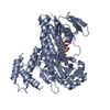
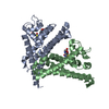
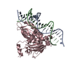
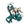
- PDB-3ks8: Crystal structure of Reston ebolavirus VP35 RNA binding domain in... -
+ Open data

- Basic information
| Entry | Database: PDB / ID: 3ks8 | ||||||
|---|---|---|---|---|---|---|---|
| Title | Crystal structure of Reston ebolavirus VP35 RNA binding domain in complex with 18bp dsRNA | ||||||
 Components Components |
| ||||||
 Keywords Keywords | VIRAL PROTEIN/RNA / ebolavirus / RNA-binding protein / protein-RNA complex / interferon / Reston / Host cytoplasm / Interferon antiviral system evasion / RNA replication / RNA-binding / Transcription / Virion / VIRAL PROTEIN-RNA complex | ||||||
| Function / homology |  Function and homology information Function and homology informationsymbiont-mediated suppression of host cytoplasmic pattern recognition receptor signaling pathway via inhibition of IKBKE activity / symbiont-mediated suppression of host cytoplasmic pattern recognition receptor signaling pathway via inhibition of IRF7 activity / virion component / symbiont-mediated suppression of host cytoplasmic pattern recognition receptor signaling pathway via inhibition of TBK1 activity / symbiont-mediated suppression of host toll-like receptor signaling pathway / host cell cytoplasm / RNA binding Similarity search - Function | ||||||
| Biological species |  Reston ebolavirus Reston ebolavirus | ||||||
| Method |  X-RAY DIFFRACTION / SYNCHROTRON / MOLECULAR REPLACEMENT / Resolution: 2.401 Å X-RAY DIFFRACTION / SYNCHROTRON / MOLECULAR REPLACEMENT / Resolution: 2.401 Å | ||||||
 Authors Authors | Kimberlin, C.R. / Bornholdt, Z.A. / Li, S. / Woods, V.L. / Macrae, I.J. / Saphire, E.O. | ||||||
 Citation Citation | Journal: Proc.Natl.Acad.Sci.USA / Year: 2009 Title: Ebolavirus VP35 uses a bimodal strategy to bind dsRNA for innate immune suppression. Authors: Kimberlin, C.R. / Bornholdt, Z.A. / Li, S. / Woods, V.L. / Macrae, I.J. / Saphire, E.O. | ||||||
| History |
|
- Structure visualization
| Structure viewer | Molecule: MolmilJmol/JSmol |
|---|
- Downloads & links
-Download
| PDBx/mmCIF format | 3ks8.cif.gz | 136.7 KB | Display | PDBx/mmCIF format |
|---|---|---|---|---|
| PDB format | pdb3ks8.ent.gz | 100.9 KB | Display | PDB format |
| PDBx/mmJSON format | 3ks8.json.gz | Tree view | PDBx/mmJSON format | |
| Others |  Other downloads Other downloads |
-Validation report
| Arichive directory | https://data.pdbj.org/pub/pdb/validation_reports/ks/3ks8ftp://data.pdbj.org/pub/pdb/validation_reports/ks/3ks8 | HTTPS FTP |
|---|
-Related structure data
| Related structure data |  3ks4SC S: Starting model for refinement C: citing same article ( |
|---|---|
| Similar structure data |


-Links
 PDBj
PDBj


- Assembly
| Deposited unit | 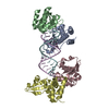
| ||||||||
|---|---|---|---|---|---|---|---|---|---|
| 1 | 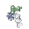
| ||||||||
| 2 | 
| ||||||||
| Unit cell |
|
-Components
| #1: RNA chain | Mass: 6056.748 Da / Num. of mol.: 1 / Source method: obtained synthetically | ||
|---|---|---|---|
| #2: RNA chain | Mass: 5455.203 Da / Num. of mol.: 1 / Source method: obtained synthetically | ||
| #3: Protein | Mass: 20569.424 Da / Num. of mol.: 4 Fragment: C-terminal RNA binding domain (UNP residues 160-329) Source method: isolated from a genetically manipulated source Source: (gene. exp.) Reston ebolavirus / Strain: Reston / Gene: REBOVgp2, VP35 / Plasmid: pET46 EK/LIC / Production host:  #4: Water | ChemComp-HOH / |  Mass: 18.015 Da / Num. of mol.: 150 / Source method: isolated from a natural source / Formula: H2O Mass: 18.015 Da / Num. of mol.: 150 / Source method: isolated from a natural source / Formula: H2O |
-Experimental details
-Experiment
| Experiment | Method: X-RAY DIFFRACTION / Number of used crystals: 1 |
|---|
- Sample preparation
| Crystal | Density Matthews: 2.46 Å3/Da / Density % sol: 49.98 % |
|---|---|
| Crystal grow | Temperature: 295.5 K / Method: vapor diffusion, hanging drop / pH: 6.5 Details: 365mM ammonium acetate, 100mM trisodium citrate, 18.5% PEG 4000, pH 6.5, VAPOR DIFFUSION, HANGING DROP, temperature 295.5K |
-Data collection
| Diffraction | Mean temperature: 100 K |
|---|---|
| Diffraction source | Source: SYNCHROTRON / Site: SSRL  / Beamline: BL11-1 / Wavelength: 0.9795 Å / Beamline: BL11-1 / Wavelength: 0.9795 Å |
| Detector | Type: MARMOSAIC 325 mm CCD / Detector: CCD / Date: May 12, 2009 |
| Radiation | Monochromator: single crystal / Protocol: SINGLE WAVELENGTH / Monochromatic (M) / Laue (L): M / Scattering type: x-ray |
| Radiation wavelength | Wavelength: 0.9795 Å / Relative weight: 1 |
| Reflection | Resolution: 2.28→37.1 Å / Num. all: 40821 / Num. obs: 33227 / % possible obs: 81.4 % / Observed criterion σ(F): 0 / Observed criterion σ(I): 0 / Redundancy: 4.3 % / Biso Wilson estimate: 42.2 Å2 / Rmerge(I) obs: 0.061 / Net I/σ(I): 23.47 |
| Reflection shell | Resolution: 2.28→2.38 Å / Redundancy: 1.43 % / Rmerge(I) obs: 0.254 / Mean I/σ(I) obs: 5.03 / Num. unique all: 1751 / % possible all: 35.3 |
- Processing
| Software |
| |||||||||||||||||||||||||||||||||||||||||||||||||||||||||||||||||||||||||||||
|---|---|---|---|---|---|---|---|---|---|---|---|---|---|---|---|---|---|---|---|---|---|---|---|---|---|---|---|---|---|---|---|---|---|---|---|---|---|---|---|---|---|---|---|---|---|---|---|---|---|---|---|---|---|---|---|---|---|---|---|---|---|---|---|---|---|---|---|---|---|---|---|---|---|---|---|---|---|---|
| Refinement | Method to determine structure: MOLECULAR REPLACEMENT Starting model: PDB entry 3KS4 Resolution: 2.401→35.117 Å / Occupancy max: 1 / Occupancy min: 0 / SU ML: 0.32 / Cross valid method: THROUGHOUT / σ(F): 1.96 / Phase error: 27.52 / Stereochemistry target values: ML
| |||||||||||||||||||||||||||||||||||||||||||||||||||||||||||||||||||||||||||||
| Solvent computation | Shrinkage radii: 0.9 Å / VDW probe radii: 1.11 Å / Solvent model: FLAT BULK SOLVENT MODEL / Bsol: 48.53 Å2 / ksol: 0.369 e/Å3 | |||||||||||||||||||||||||||||||||||||||||||||||||||||||||||||||||||||||||||||
| Displacement parameters | Biso max: 128.04 Å2 / Biso mean: 42.743 Å2 / Biso min: 10.07 Å2
| |||||||||||||||||||||||||||||||||||||||||||||||||||||||||||||||||||||||||||||
| Refinement step | Cycle: LAST / Resolution: 2.401→35.117 Å
| |||||||||||||||||||||||||||||||||||||||||||||||||||||||||||||||||||||||||||||
| Refine LS restraints |
| |||||||||||||||||||||||||||||||||||||||||||||||||||||||||||||||||||||||||||||
| LS refinement shell | Refine-ID: X-RAY DIFFRACTION
|
