 Movie
Movie Controller

[English] 日本語
 Yorodumi
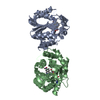
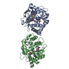
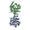
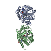
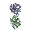
- PDB-3kqt: Crystal Structure of hPNMT in Complex AdoHcy and 2-Amino-1-methyl... -
+ Open data

- Basic information
| Entry | Database: PDB / ID: 3kqt | ||||||
|---|---|---|---|---|---|---|---|
| Title | Crystal Structure of hPNMT in Complex AdoHcy and 2-Amino-1-methylbenzimidazole | ||||||
 Components Components | Phenylethanolamine N-methyltransferase | ||||||
 Keywords Keywords | TRANSFERASE / methyltransferase / fragment screening / Catecholamine biosynthesis / S-adenosyl-L-methionine | ||||||
| Function / homology |  Function and homology information Function and homology informationphenylethanolamine N-methyltransferase / phenylethanolamine N-methyltransferase activity / epinephrine biosynthetic process / Catecholamine biosynthesis / catecholamine biosynthetic process / methylation / cytosol Similarity search - Function | ||||||
| Biological species |  Homo sapiens (human) Homo sapiens (human) | ||||||
| Method |  X-RAY DIFFRACTION / SYNCHROTRON / Resolution: 2.399 Å X-RAY DIFFRACTION / SYNCHROTRON / Resolution: 2.399 Å | ||||||
 Authors Authors | Drinkwater, N. / Martin, J.L. | ||||||
 Citation Citation | Journal: Biochem.J. / Year: 2010 Title: Fragment-based screening by X-ray crystallography, MS and isothermal titration calorimetry to identify PNMT (phenylethanolamine N-methyltransferase) inhibitors. Authors: Drinkwater, N. / Vu, H. / Lovell, K.M. / Criscione, K.R. / Collins, B.M. / Prisinzano, T.E. / Poulsen, S.A. / McLeish, M.J. / Grunewald, G.L. / Martin, J.L. | ||||||
| History |
|
- Structure visualization
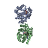
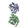
| Structure viewer | Molecule: MolmilJmol/JSmol |
|---|
- Downloads & links
-Download
| PDBx/mmCIF format | 3kqt.cif.gz | 232.6 KB | Display | PDBx/mmCIF format |
|---|---|---|---|---|
| PDB format | pdb3kqt.ent.gz | 187.2 KB | Display | PDB format |
| PDBx/mmJSON format | 3kqt.json.gz | Tree view | PDBx/mmJSON format | |
| Others |  Other downloads Other downloads |
-Validation report
| Arichive directory | https://data.pdbj.org/pub/pdb/validation_reports/kq/3kqtftp://data.pdbj.org/pub/pdb/validation_reports/kq/3kqt | HTTPS FTP |
|---|
-Related structure data
| Related structure data | 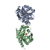 3kpjC 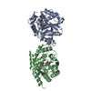 3kpuC  3kpvC  3kpwC 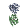 3kpyC  3kqmC  3kqoC  3kqpC  3kqqC  3kqsC 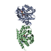 3kqvC 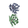 3kqwC  3kqyC  3kr0C 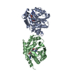 3kr1C  3kr2C 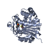 1hnnS C: citing same article ( S: Starting model for refinement |
|---|---|
| Similar structure data |


-Links
 PDBj
PDBj- Assembly
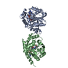
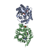
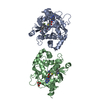
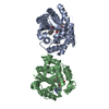
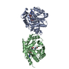
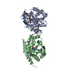
| Deposited unit | 
| ||||||||
|---|---|---|---|---|---|---|---|---|---|
| 1 |
| ||||||||
| Unit cell |
|
-Components
| #1: Protein | Mass: 31845.967 Da / Num. of mol.: 2 Source method: isolated from a genetically manipulated source Source: (gene. exp.) Homo sapiens (human) / Gene: PNMT, PENT / Plasmid: pET17 PNMT-His / Production host:  References: UniProt: P11086, phenylethanolamine N-methyltransferase #2: Chemical |   Mass: 384.411 Da / Num. of mol.: 2 / Source method: obtained synthetically / Formula: C14H20N6O5S Mass: 384.411 Da / Num. of mol.: 2 / Source method: obtained synthetically / Formula: C14H20N6O5S#3: Chemical | 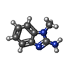  Mass: 147.177 Da / Num. of mol.: 2 / Source method: obtained synthetically / Formula: C8H9N3 Mass: 147.177 Da / Num. of mol.: 2 / Source method: obtained synthetically / Formula: C8H9N3#4: Water | ChemComp-HOH / |  Mass: 18.015 Da / Num. of mol.: 277 / Source method: isolated from a natural source / Formula: H2O Mass: 18.015 Da / Num. of mol.: 277 / Source method: isolated from a natural source / Formula: H2OHas protein modification | Y | |
|---|
-Experimental details
-Experiment
| Experiment | Method: X-RAY DIFFRACTION / Number of used crystals: 1 |
|---|
- Sample preparation
| Crystal | Density Matthews: 3.26 Å3/Da / Density % sol: 62.25 % |
|---|---|
| Crystal grow | Temperature: 298 K / Method: vapor diffusion, hanging drop / pH: 5.8 Details: PEG6K, LiCl, cacodylate, pH 5.8, vapor diffusion, hanging drop, temperature 298K |
-Data collection
| Diffraction | Mean temperature: 100 K | ||||||||||||||||||||||||||||||||||||||||||||||||||||||||||||||||||
|---|---|---|---|---|---|---|---|---|---|---|---|---|---|---|---|---|---|---|---|---|---|---|---|---|---|---|---|---|---|---|---|---|---|---|---|---|---|---|---|---|---|---|---|---|---|---|---|---|---|---|---|---|---|---|---|---|---|---|---|---|---|---|---|---|---|---|---|
| Diffraction source | Source: SYNCHROTRON / Site: Australian Synchrotron  / Beamline: MX1 / Wavelength: 0.95667 Å / Beamline: MX1 / Wavelength: 0.95667 Å | ||||||||||||||||||||||||||||||||||||||||||||||||||||||||||||||||||
| Detector | Type: MAR CCD 165 mm / Detector: CCD / Date: Nov 30, 2007 | ||||||||||||||||||||||||||||||||||||||||||||||||||||||||||||||||||
| Radiation | Protocol: SINGLE WAVELENGTH / Monochromatic (M) / Laue (L): M / Scattering type: x-ray | ||||||||||||||||||||||||||||||||||||||||||||||||||||||||||||||||||
| Radiation wavelength | Wavelength: 0.95667 Å / Relative weight: 1 | ||||||||||||||||||||||||||||||||||||||||||||||||||||||||||||||||||
| Reflection | Resolution: 2.4→50 Å / Num. obs: 33746 / % possible obs: 99.7 % / Redundancy: 9.4 % / Rmerge(I) obs: 0.077 / Χ2: 1.036 / Net I/σ(I): 9.1 | ||||||||||||||||||||||||||||||||||||||||||||||||||||||||||||||||||
| Reflection shell |
|
- Processing
| Software |
| |||||||||||||||||||||||||||||||||||||||||||||||||||||||||||||||||||||||||||||||||||||||||||
|---|---|---|---|---|---|---|---|---|---|---|---|---|---|---|---|---|---|---|---|---|---|---|---|---|---|---|---|---|---|---|---|---|---|---|---|---|---|---|---|---|---|---|---|---|---|---|---|---|---|---|---|---|---|---|---|---|---|---|---|---|---|---|---|---|---|---|---|---|---|---|---|---|---|---|---|---|---|---|---|---|---|---|---|---|---|---|---|---|---|---|---|---|
| Refinement | Starting model: PDB entry 1HNN Resolution: 2.399→19.486 Å / Occupancy max: 1 / Occupancy min: 0.5 / FOM work R set: 0.821 / SU ML: 0.34 / Cross valid method: THROUGHOUT / σ(F): 1.33 / Stereochemistry target values: ML
| |||||||||||||||||||||||||||||||||||||||||||||||||||||||||||||||||||||||||||||||||||||||||||
| Solvent computation | Shrinkage radii: 0.9 Å / VDW probe radii: 1.11 Å / Solvent model: FLAT BULK SOLVENT MODEL / Bsol: 51.945 Å2 / ksol: 0.328 e/Å3 | |||||||||||||||||||||||||||||||||||||||||||||||||||||||||||||||||||||||||||||||||||||||||||
| Displacement parameters | Biso max: 132.66 Å2 / Biso mean: 47.986 Å2 / Biso min: 19.7 Å2
| |||||||||||||||||||||||||||||||||||||||||||||||||||||||||||||||||||||||||||||||||||||||||||
| Refinement step | Cycle: LAST / Resolution: 2.399→19.486 Å
| |||||||||||||||||||||||||||||||||||||||||||||||||||||||||||||||||||||||||||||||||||||||||||
| Refine LS restraints |
| |||||||||||||||||||||||||||||||||||||||||||||||||||||||||||||||||||||||||||||||||||||||||||
| LS refinement shell | Refine-ID: X-RAY DIFFRACTION / Total num. of bins used: 12
| |||||||||||||||||||||||||||||||||||||||||||||||||||||||||||||||||||||||||||||||||||||||||||
| Refinement TLS params. | Method: refined / Origin x: 23.9574 Å / Origin y: 51.0101 Å / Origin z: -5.9121 Å
| |||||||||||||||||||||||||||||||||||||||||||||||||||||||||||||||||||||||||||||||||||||||||||
| Refinement TLS group |
|
