 Movie
Movie Controller

[English] 日本語
 Yorodumi
- PDB-1hnn: CRYSTAL STRUCTURE OF HUMAN PNMT COMPLEXED WITH SK&F 29661 AND ADO... -
+ Open data

- Basic information
| Entry | Database: PDB / ID: 1hnn | ||||||
|---|---|---|---|---|---|---|---|
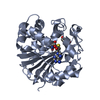
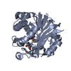
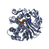
| Title | CRYSTAL STRUCTURE OF HUMAN PNMT COMPLEXED WITH SK&F 29661 AND ADOHCY(SAH) | ||||||
 Components Components | PHENYLETHANOLAMINE N-METHYLTRANSFERASE | ||||||
 Keywords Keywords | TRANSFERASE / methyltransferase / adrenaline synthesis / S-adenosyl methionine | ||||||
| Function / homology |  Function and homology information Function and homology informationphenylethanolamine N-methyltransferase / phenylethanolamine N-methyltransferase activity / epinephrine biosynthetic process / Catecholamine biosynthesis / catecholamine biosynthetic process / methylation / cytosol Similarity search - Function | ||||||
| Biological species |  Homo sapiens (human) Homo sapiens (human) | ||||||
| Method |  X-RAY DIFFRACTION / MIR / Resolution: 2.4 Å X-RAY DIFFRACTION / MIR / Resolution: 2.4 Å | ||||||
 Authors Authors | Martin, J.L. / Begun, J. / McLeish, M.J. / Caine, J.M. / Grunewald, G.L. | ||||||
 Citation Citation | Journal: Structure / Year: 2001 Title: Getting the adrenaline going: crystal structure of the adrenaline-synthesizing enzyme PNMT. Authors: Martin, J.L. / Begun, J. / McLeish, M.J. / Caine, J.M. / Grunewald, G.L. | ||||||
| History |
|
- Structure visualization
| Structure viewer | Molecule: MolmilJmol/JSmol |
|---|
- Downloads & links
-Download
| PDBx/mmCIF format | 1hnn.cif.gz | 119.5 KB | Display | PDBx/mmCIF format |
|---|---|---|---|---|
| PDB format | pdb1hnn.ent.gz | 92.3 KB | Display | PDB format |
| PDBx/mmJSON format | 1hnn.json.gz | Tree view | PDBx/mmJSON format | |
| Others |  Other downloads Other downloads |
-Validation report
| Arichive directory | https://data.pdbj.org/pub/pdb/validation_reports/hn/1hnnftp://data.pdbj.org/pub/pdb/validation_reports/hn/1hnn | HTTPS FTP |
|---|
-Related structure data
| Similar structure data |
|---|


-Links
 PDBj
PDBj- Assembly
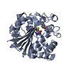
| Deposited unit | 
| ||||||||
|---|---|---|---|---|---|---|---|---|---|
| 1 | 
| ||||||||
| 2 | 
| ||||||||
| Unit cell |
|
-Components
| #1: Protein | Mass: 30887.982 Da / Num. of mol.: 2 Source method: isolated from a genetically manipulated source Source: (gene. exp.) Homo sapiens (human) / Gene: PNMT / Production host:  References: UniProt: P11086, phenylethanolamine N-methyltransferase #2: Chemical |   Type: L-peptide linking / Mass: 384.411 Da / Num. of mol.: 2 / Source method: obtained synthetically / Formula: C14H20N6O5S Type: L-peptide linking / Mass: 384.411 Da / Num. of mol.: 2 / Source method: obtained synthetically / Formula: C14H20N6O5S#3: Chemical |   Mass: 212.269 Da / Num. of mol.: 2 / Source method: obtained synthetically / Formula: C9H12N2O2S Mass: 212.269 Da / Num. of mol.: 2 / Source method: obtained synthetically / Formula: C9H12N2O2S#4: Water | ChemComp-HOH / |  Mass: 18.015 Da / Num. of mol.: 126 / Source method: isolated from a natural source / Formula: H2O Mass: 18.015 Da / Num. of mol.: 126 / Source method: isolated from a natural source / Formula: H2O |
|---|
-Experimental details
-Experiment
| Experiment | Method: X-RAY DIFFRACTION / Number of used crystals: 1 |
|---|
- Sample preparation
| Crystal | Density Matthews: 3.38 Å3/Da / Density % sol: 63.6 % | |||||||||||||||
|---|---|---|---|---|---|---|---|---|---|---|---|---|---|---|---|---|
| Crystal grow | Temperature: 293 K / Method: vapor diffusion, hanging drop / pH: 6.5 Details: PEG 6K, pH 6.5, VAPOR DIFFUSION, HANGING DROP, temperature 293K | |||||||||||||||
| Crystal grow | *PLUS | |||||||||||||||
| Components of the solutions | *PLUS
|
-Data collection
| Diffraction | Mean temperature: 100 K |
|---|---|
| Diffraction source | Source: ROTATING ANODE / Type: RIGAKU RU200 / Wavelength: 1.5418 Å |
| Detector | Type: RIGAKU RAXIS IIC / Detector: IMAGE PLATE / Date: Feb 14, 1997 / Details: Yale mirrors |
| Radiation | Monochromator: Ni filter / Protocol: SINGLE WAVELENGTH / Monochromatic (M) / Laue (L): M / Scattering type: x-ray |
| Radiation wavelength | Wavelength: 1.5418 Å / Relative weight: 1 |
| Reflection | Resolution: 2.3→38.48 Å / Num. all: 38545 / Num. obs: 34845 / % possible obs: 90.4 % / Observed criterion σ(F): 0 / Observed criterion σ(I): 0 / Redundancy: 4.7 % / Biso Wilson estimate: 43.5 Å2 / Rmerge(I) obs: 0.051 / Net I/σ(I): 18.9 |
| Reflection shell | Resolution: 2.3→2.38 Å / Redundancy: 1.6 % / Rmerge(I) obs: 0.274 / Mean I/σ(I) obs: 2.1 / % possible all: 54 |
| Reflection | *PLUS Lowest resolution: 50 Å |
| Reflection shell | *PLUS % possible obs: 54 % |
- Processing
| Software |
| ||||||||||||||||||||||||||||||||||||||||
|---|---|---|---|---|---|---|---|---|---|---|---|---|---|---|---|---|---|---|---|---|---|---|---|---|---|---|---|---|---|---|---|---|---|---|---|---|---|---|---|---|---|
| Refinement | Method to determine structure: MIR / Resolution: 2.4→38.48 Å / Rfactor Rfree error: 0.005 / Data cutoff high absF: 444544.74 / Data cutoff low absF: 0 / Isotropic thermal model: RESTRAINED / Cross valid method: THROUGHOUT / σ(F): 0 / σ(I): 0 / Stereochemistry target values: Engh and Huber
| ||||||||||||||||||||||||||||||||||||||||
| Solvent computation | Solvent model: FLAT MODEL / Bsol: 30.19 Å2 / ksol: 0.319 e/Å3 | ||||||||||||||||||||||||||||||||||||||||
| Displacement parameters | Biso mean: 45 Å2
| ||||||||||||||||||||||||||||||||||||||||
| Refine analyze |
| ||||||||||||||||||||||||||||||||||||||||
| Refinement step | Cycle: LAST / Resolution: 2.4→38.48 Å
| ||||||||||||||||||||||||||||||||||||||||
| Refine LS restraints |
| ||||||||||||||||||||||||||||||||||||||||
| LS refinement shell | Resolution: 2.4→2.55 Å / Rfactor Rfree error: 0.017 / Total num. of bins used: 6
| ||||||||||||||||||||||||||||||||||||||||
| Xplor file |
| ||||||||||||||||||||||||||||||||||||||||
| Software | *PLUS Name: CNS / Version: 0.9 / Classification: refinement | ||||||||||||||||||||||||||||||||||||||||
| Refinement | *PLUS Lowest resolution: 50 Å / σ(F): 0 / % reflection Rfree: 10 % / Rfactor obs: 0.231 / Rfactor Rfree: 0.272 / Rfactor Rwork: 0.23 | ||||||||||||||||||||||||||||||||||||||||
| Solvent computation | *PLUS | ||||||||||||||||||||||||||||||||||||||||
| Displacement parameters | *PLUS Biso mean: 45 Å2 | ||||||||||||||||||||||||||||||||||||||||
| Refine LS restraints | *PLUS
| ||||||||||||||||||||||||||||||||||||||||
| LS refinement shell | *PLUS Rfactor Rfree: 0.367 / % reflection Rfree: 10.4 % / Rfactor Rwork: 0.352 |
