 Movie
Movie Controller

[English] 日本語
 Yorodumi
- PDB-3hca: Crystal Structure of E185Q hPNMT in Complex With Octopamine and AdoHcy -
+ Open data

- Basic information
| Entry | Database: PDB / ID: 3hca | ||||||
|---|---|---|---|---|---|---|---|
| Title | Crystal Structure of E185Q hPNMT in Complex With Octopamine and AdoHcy | ||||||
 Components Components | Phenylethanolamine N-methyltransferase | ||||||
 Keywords Keywords | TRANSFERASE / methyltransferase / Catecholamine biosynthesis / Polymorphism / S-adenosyl-L-methionine | ||||||
| Function / homology |  Function and homology information Function and homology informationphenylethanolamine N-methyltransferase / phenylethanolamine N-methyltransferase activity / epinephrine biosynthetic process / Catecholamine biosynthesis / catecholamine biosynthetic process / methylation / cytosol Similarity search - Function | ||||||
| Biological species |  Homo sapiens (human) Homo sapiens (human) | ||||||
| Method |  X-RAY DIFFRACTION / FOURIER SYNTHESIS / Resolution: 2.4 Å X-RAY DIFFRACTION / FOURIER SYNTHESIS / Resolution: 2.4 Å | ||||||
 Authors Authors | Drinkwater, N. / Martin, J.L. | ||||||
 Citation Citation | Journal: Biochem.J. / Year: 2009 Title: Molecular recognition of physiological substrate noradrenaline by the adrenaline-synthesizing enzyme PNMT and factors influencing its methyltransferase activity. Authors: Drinkwater, N. / Gee, C.L. / Puri, M. / Criscione, K.R. / McLeish, M.J. / Grunewald, G.L. / Martin, J.L. | ||||||
| History |
|
- Structure visualization
| Structure viewer | Molecule: MolmilJmol/JSmol |
|---|
- Downloads & links
-Download
| PDBx/mmCIF format | 3hca.cif.gz | 232.8 KB | Display | PDBx/mmCIF format |
|---|---|---|---|---|
| PDB format | pdb3hca.ent.gz | 188.1 KB | Display | PDB format |
| PDBx/mmJSON format | 3hca.json.gz | Tree view | PDBx/mmJSON format | |
| Others |  Other downloads Other downloads |
-Validation report
| Arichive directory | https://data.pdbj.org/pub/pdb/validation_reports/hc/3hcaftp://data.pdbj.org/pub/pdb/validation_reports/hc/3hca | HTTPS FTP |
|---|
-Related structure data
| Related structure data |  3hcbC  3hccC  3hcdC  3hceC  3hcfC  1hnnS S: Starting model for refinement C: citing same article ( |
|---|---|
| Similar structure data |


-Links
 PDBj
PDBj- Assembly
| Deposited unit | 
| ||||||||
|---|---|---|---|---|---|---|---|---|---|
| 1 | 
| ||||||||
| 2 | 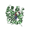
| ||||||||
| 3 |
| ||||||||
| Unit cell |
|
-Components
| #1: Protein | Mass: 31844.982 Da / Num. of mol.: 2 / Mutation: E185Q Source method: isolated from a genetically manipulated source Source: (gene. exp.) Homo sapiens (human) / Gene: PNMT, PENT / Plasmid: pET17 PNMT-His / Production host:  References: UniProt: P11086, phenylethanolamine N-methyltransferase #2: Chemical |   Mass: 384.411 Da / Num. of mol.: 2 / Source method: obtained synthetically / Formula: C14H20N6O5S Mass: 384.411 Da / Num. of mol.: 2 / Source method: obtained synthetically / Formula: C14H20N6O5S#3: Chemical |   Mass: 153.178 Da / Num. of mol.: 2 / Source method: obtained synthetically / Formula: C8H11NO2 Mass: 153.178 Da / Num. of mol.: 2 / Source method: obtained synthetically / Formula: C8H11NO2#4: Chemical | ChemComp-EDO / |   Mass: 62.068 Da / Num. of mol.: 1 / Source method: obtained synthetically / Formula: C2H6O2 Mass: 62.068 Da / Num. of mol.: 1 / Source method: obtained synthetically / Formula: C2H6O2#5: Water | ChemComp-HOH / |  Mass: 18.015 Da / Num. of mol.: 310 / Source method: isolated from a natural source / Formula: H2O Mass: 18.015 Da / Num. of mol.: 310 / Source method: isolated from a natural source / Formula: H2OHas protein modification | Y | |
|---|
-Experimental details
-Experiment
| Experiment | Method: X-RAY DIFFRACTION / Number of used crystals: 1 |
|---|
- Sample preparation
| Crystal | Density Matthews: 3.27 Å3/Da / Density % sol: 62.43 % |
|---|---|
| Crystal grow | Temperature: 298 K / Method: vapor diffusion, hanging drop / pH: 5.8 Details: PEG6K, LiCl, cacodylate, pH 5.8, vapor diffusion, hanging drop, temperature 298K |
-Data collection
| Diffraction | Mean temperature: 100 K |
|---|---|
| Diffraction source | Source: ROTATING ANODE / Type: RIGAKU FR-E SUPERBRIGHT / Wavelength: 1.5418 Å |
| Detector | Type: RIGAKU SATURN 944 / Detector: CCD / Date: Oct 8, 2007 / Details: OSMIC VARI-MAX HF |
| Radiation | Monochromator: OSMIC VARI-MAX HF / Protocol: SINGLE WAVELENGTH / Monochromatic (M) / Laue (L): M / Scattering type: x-ray |
| Radiation wavelength | Wavelength: 1.5418 Å / Relative weight: 1 |
| Reflection | Resolution: 2.4→42.11 Å / Num. obs: 30563 / Redundancy: 6.56 % / Biso Wilson estimate: 41.62 Å2 / Rmerge(I) obs: 0.069 / Net I/σ(I): 17 |
| Reflection shell | Resolution: 2.4→2.49 Å / Redundancy: 3.21 % / Rmerge(I) obs: 0.55 / Mean I/σ(I) obs: 1.5 |
- Processing
| Software |
| |||||||||||||||||||||||||||||||||||||||||||||||||||||||||||||||||||||||||||||
|---|---|---|---|---|---|---|---|---|---|---|---|---|---|---|---|---|---|---|---|---|---|---|---|---|---|---|---|---|---|---|---|---|---|---|---|---|---|---|---|---|---|---|---|---|---|---|---|---|---|---|---|---|---|---|---|---|---|---|---|---|---|---|---|---|---|---|---|---|---|---|---|---|---|---|---|---|---|---|
| Refinement | Method to determine structure: FOURIER SYNTHESIS Starting model: 1HNN Resolution: 2.4→42.105 Å / Occupancy max: 1 / Occupancy min: 0 / SU ML: 0.3 / Isotropic thermal model: RESTRAINED / Cross valid method: THROUGHOUT / σ(F): 0.01 / Stereochemistry target values: ML
| |||||||||||||||||||||||||||||||||||||||||||||||||||||||||||||||||||||||||||||
| Solvent computation | Shrinkage radii: 0.9 Å / VDW probe radii: 1.11 Å / Solvent model: FLAT BULK SOLVENT MODEL / Bsol: 37.319 Å2 / ksol: 0.309 e/Å3 | |||||||||||||||||||||||||||||||||||||||||||||||||||||||||||||||||||||||||||||
| Displacement parameters | Biso max: 112.18 Å2 / Biso mean: 44.104 Å2 / Biso min: 16.04 Å2
| |||||||||||||||||||||||||||||||||||||||||||||||||||||||||||||||||||||||||||||
| Refinement step | Cycle: LAST / Resolution: 2.4→42.105 Å
| |||||||||||||||||||||||||||||||||||||||||||||||||||||||||||||||||||||||||||||
| Refine LS restraints |
| |||||||||||||||||||||||||||||||||||||||||||||||||||||||||||||||||||||||||||||
| LS refinement shell | Refine-ID: X-RAY DIFFRACTION / Total num. of bins used: 10
| |||||||||||||||||||||||||||||||||||||||||||||||||||||||||||||||||||||||||||||
| Refinement TLS params. | Method: refined / Origin x: 24.103 Å / Origin y: 51.3065 Å / Origin z: -5.7897 Å
| |||||||||||||||||||||||||||||||||||||||||||||||||||||||||||||||||||||||||||||
| Refinement TLS group |
|
