 Movie
Movie Controller

[English] 日本語
 Yorodumi
- PDB-2g70: Structure of human PNMT in complex with inhibitor 3-hydroxymethyl... -
+ Open data

- Basic information
| Entry | Database: PDB / ID: 2g70 | ||||||
|---|---|---|---|---|---|---|---|
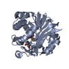
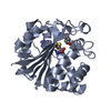
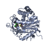
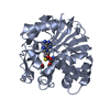
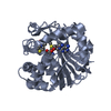
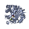
| Title | Structure of human PNMT in complex with inhibitor 3-hydroxymethyl-7-nitro-THIQ and AdoMet (SAM) | ||||||
 Components Components | Phenylethanolamine N-methyltransferase | ||||||
 Keywords Keywords | TRANSFERASE / methyltransferase | ||||||
| Function / homology |  Function and homology information Function and homology informationphenylethanolamine N-methyltransferase / phenylethanolamine N-methyltransferase activity / epinephrine biosynthetic process / Catecholamine biosynthesis / catecholamine biosynthetic process / methylation / cytosol Similarity search - Function | ||||||
| Biological species |  Homo sapiens (human) Homo sapiens (human) | ||||||
| Method |  X-RAY DIFFRACTION / FOURIER SYNTHESIS / Resolution: 2.4 Å X-RAY DIFFRACTION / FOURIER SYNTHESIS / Resolution: 2.4 Å | ||||||
 Authors Authors | Tyndall, J.D.A. / Gee, C.L. / Martin, J.L. | ||||||
 Citation Citation | Journal: J.Med.Chem. / Year: 2007 Title: Enzyme Adaptation to Inhibitor Binding: A Cryptic Binding Site in Phenylethanolamine N-Methyltransferase Authors: Gee, C.L. / Drinkwater, N. / Tyndall, J.D.A. / Grunewald, G.L. / Wu, Q. / McLeish, M.J. / Martin, J.L. | ||||||
| History |
|
- Structure visualization
| Structure viewer | Molecule: MolmilJmol/JSmol |
|---|
- Downloads & links
-Download
| PDBx/mmCIF format | 2g70.cif.gz | 119.4 KB | Display | PDBx/mmCIF format |
|---|---|---|---|---|
| PDB format | pdb2g70.ent.gz | 93 KB | Display | PDB format |
| PDBx/mmJSON format | 2g70.json.gz | Tree view | PDBx/mmJSON format | |
| Others |  Other downloads Other downloads |
-Validation report
| Arichive directory | https://data.pdbj.org/pub/pdb/validation_reports/g7/2g70ftp://data.pdbj.org/pub/pdb/validation_reports/g7/2g70 | HTTPS FTP |
|---|
-Related structure data
| Related structure data |  2g71C  2g72C  2obfC  2onyC  2onzC  2opbC 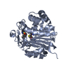 1hnnS S: Starting model for refinement C: citing same article ( |
|---|---|
| Similar structure data |


-Links
 PDBj
PDBj
- Assembly
| Deposited unit | 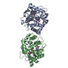
| ||||||||
|---|---|---|---|---|---|---|---|---|---|
| 1 | 
| ||||||||
| 2 | 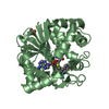
| ||||||||
| 3 |
| ||||||||
| Unit cell |
|
-Components
| #1: Protein | Mass: 31845.967 Da / Num. of mol.: 2 Source method: isolated from a genetically manipulated source Source: (gene. exp.) Homo sapiens (human) / Plasmid: PNMT-His / Production host:  References: UniProt: P11086, phenylethanolamine N-methyltransferase #2: Chemical |   Mass: 398.437 Da / Num. of mol.: 2 / Source method: obtained synthetically / Formula: C15H22N6O5S Mass: 398.437 Da / Num. of mol.: 2 / Source method: obtained synthetically / Formula: C15H22N6O5S#3: Chemical |   Mass: 208.214 Da / Num. of mol.: 3 / Source method: obtained synthetically / Formula: C10H12N2O3 Mass: 208.214 Da / Num. of mol.: 3 / Source method: obtained synthetically / Formula: C10H12N2O3#4: Chemical | ChemComp-PO4 / |   Mass: 94.971 Da / Num. of mol.: 1 / Source method: obtained synthetically / Formula: PO4 Mass: 94.971 Da / Num. of mol.: 1 / Source method: obtained synthetically / Formula: PO4#5: Water | ChemComp-HOH / |  Mass: 18.015 Da / Num. of mol.: 81 / Source method: isolated from a natural source / Formula: H2O Mass: 18.015 Da / Num. of mol.: 81 / Source method: isolated from a natural source / Formula: H2OHas protein modification | Y | |
|---|
-Experimental details
-Experiment
| Experiment | Method: X-RAY DIFFRACTION / Number of used crystals: 1 |
|---|
- Sample preparation
| Crystal | Density Matthews: 3.27 Å3/Da / Density % sol: 62.4 % |
|---|---|
| Crystal grow | Temperature: 293 K / Method: vapor diffusion, hanging drop / pH: 5.3 Details: 0.6M ammonium phosphate, 0.1M Sodium citrate, pH 5.3, VAPOR DIFFUSION, HANGING DROP, temperature 293K |
-Data collection
| Diffraction | Mean temperature: 100 K |
|---|---|
| Diffraction source | Source: ROTATING ANODE / Type: RIGAKU FR-E+ DW / Wavelength: 1.5418 Å |
| Detector | Type: RIGAKU RAXIS IV++ / Detector: IMAGE PLATE / Date: Jul 1, 2003 |
| Radiation | Monochromator: HiRes2 / Protocol: SINGLE WAVELENGTH / Monochromatic (M) / Laue (L): M / Scattering type: x-ray |
| Radiation wavelength | Wavelength: 1.5418 Å / Relative weight: 1 |
| Reflection | Resolution: 2.4→29.8 Å / Num. obs: 33540 / % possible obs: 99 % / Redundancy: 7.07 % / Biso Wilson estimate: 30.9 Å2 / Rmerge(I) obs: 0.074 / Χ2: 0.97 / Net I/σ(I): 12.7 / Scaling rejects: 1792 |
| Reflection shell | Resolution: 2.4→2.49 Å / % possible obs: 97.9 % / Redundancy: 7.25 % / Rmerge(I) obs: 0.559 / Mean I/σ(I) obs: 3.6 / Num. measured all: 23577 / Num. unique all: 3234 / Num. unique obs: 3249 / Χ2: 1.08 / % possible all: 97.9 |
- Processing
| Software |
| ||||||||||||||||||||||||||||
|---|---|---|---|---|---|---|---|---|---|---|---|---|---|---|---|---|---|---|---|---|---|---|---|---|---|---|---|---|---|
| Refinement | Method to determine structure: FOURIER SYNTHESIS Starting model: 1HNN Resolution: 2.4→29.8 Å / Isotropic thermal model: Restrained / Cross valid method: THROUGHOUT / σ(F): 0 / Stereochemistry target values: Engh & Huber
| ||||||||||||||||||||||||||||
| Solvent computation | Bsol: 40.432 Å2 | ||||||||||||||||||||||||||||
| Displacement parameters | Biso mean: 60.036 Å2
| ||||||||||||||||||||||||||||
| Refine analyze |
| ||||||||||||||||||||||||||||
| Refinement step | Cycle: LAST / Resolution: 2.4→29.8 Å
| ||||||||||||||||||||||||||||
| Refine LS restraints |
| ||||||||||||||||||||||||||||
| LS refinement shell | Resolution: 2.4→2.49 Å / Rfactor Rfree error: 0.025
| ||||||||||||||||||||||||||||
| Xplor file |
|
