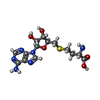
+ Open data
- Basic information
| Entry | Database: PDB / ID: 3kpj | ||||||
|---|---|---|---|---|---|---|---|
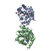
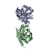
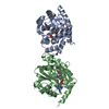
| Title | Crystal Structure of hPNMT in Complex AdoHcy and Bound Phosphate | ||||||
 Components Components | Phenylethanolamine N-methyltransferase | ||||||
 Keywords Keywords | TRANSFERASE / methyltransferase / fragment screening / Catecholamine biosynthesis / S-adenosyl-L-methionine | ||||||
| Function / homology |  Function and homology information Function and homology informationphenylethanolamine N-methyltransferase / phenylethanolamine N-methyltransferase activity / epinephrine biosynthetic process / Catecholamine biosynthesis / catecholamine biosynthetic process / methylation / cytosol Similarity search - Function | ||||||
| Biological species |  Homo sapiens (human) Homo sapiens (human) | ||||||
| Method |  X-RAY DIFFRACTION / X-RAY DIFFRACTION /  FOURIER SYNTHESIS / Resolution: 2.5 Å FOURIER SYNTHESIS / Resolution: 2.5 Å | ||||||
 Authors Authors | Drinkwater, N. / Martin, J.L. | ||||||
 Citation Citation |  Journal: Biochem.J. / Year: 2010 Journal: Biochem.J. / Year: 2010Title: Fragment-based screening by X-ray crystallography, MS and isothermal titration calorimetry to identify PNMT (phenylethanolamine N-methyltransferase) inhibitors. Authors: Drinkwater, N. / Vu, H. / Lovell, K.M. / Criscione, K.R. / Collins, B.M. / Prisinzano, T.E. / Poulsen, S.A. / McLeish, M.J. / Grunewald, G.L. / Martin, J.L. | ||||||
| History |
|
- Structure visualization
| Structure viewer | Molecule:  Molmil Molmil Jmol/JSmol Jmol/JSmol |
|---|
- Downloads & links
- Download
| PDBx/mmCIF format |  3kpj.cif.gz 3kpj.cif.gz | 225.4 KB | Display |  PDBx/mmCIF format PDBx/mmCIF format |
|---|---|---|---|---|
| PDB format |  pdb3kpj.ent.gz pdb3kpj.ent.gz | 183.7 KB | Display |  PDB format PDB format |
| PDBx/mmJSON format |  3kpj.json.gz 3kpj.json.gz | Tree view |  PDBx/mmJSON format PDBx/mmJSON format | |
| Others |  Other downloads Other downloads |
-Validation report
| Arichive directory |  https://data.pdbj.org/pub/pdb/validation_reports/kp/3kpj https://data.pdbj.org/pub/pdb/validation_reports/kp/3kpj ftp://data.pdbj.org/pub/pdb/validation_reports/kp/3kpj ftp://data.pdbj.org/pub/pdb/validation_reports/kp/3kpj | HTTPS FTP |
|---|
-Related structure data
| Related structure data |  3kpuC  3kpvC  3kpwC  3kpyC  3kqmC  3kqoC  3kqpC 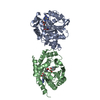 3kqqC 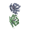 3kqsC 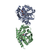 3kqtC 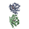 3kqvC  3kqwC  3kqyC  3kr0C  3kr1C  3kr2C  1hnnS C: citing same article ( S: Starting model for refinement |
|---|---|
| Similar structure data |
- Links
Links
- Assembly
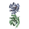
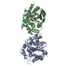
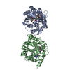
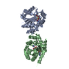
| Deposited unit | 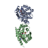
| ||||||||
|---|---|---|---|---|---|---|---|---|---|
| 1 |
| ||||||||
| Unit cell |
|
- Components
| #1: Protein | Mass: 31845.967 Da / Num. of mol.: 2 Source method: isolated from a genetically manipulated source Source: (gene. exp.)  Homo sapiens (human) / Gene: PENT, PNMT / Plasmid: pET17 PNMT-His / Production host: Homo sapiens (human) / Gene: PENT, PNMT / Plasmid: pET17 PNMT-His / Production host:  References: UniProt: P11086, phenylethanolamine N-methyltransferase #2: Chemical | #3: Chemical | #4: Water | ChemComp-HOH / | Has protein modification | Y | |
|---|
-Experimental details
-Experiment
| Experiment | Method:  X-RAY DIFFRACTION / Number of used crystals: 1 X-RAY DIFFRACTION / Number of used crystals: 1 |
|---|
- Sample preparation
| Crystal | Density Matthews: 3.27 Å3/Da / Density % sol: 62.39 % |
|---|---|
| Crystal grow | Temperature: 298 K / Method: vapor diffusion, hanging drop / pH: 5.8 Details: PEG6K, LiCl, cacodylate, pH 5.8, vapor diffusion, hanging drop, temperature 298K |
-Data collection
| Diffraction | Mean temperature: 100 K |
|---|---|
| Diffraction source | Source:  ROTATING ANODE / Type: RIGAKU FR-E / Wavelength: 1.5418 Å ROTATING ANODE / Type: RIGAKU FR-E / Wavelength: 1.5418 Å |
| Detector | Type: RIGAKU RAXIS / Detector: IMAGE PLATE / Date: Aug 3, 2007 |
| Radiation | Monochromator: HiRes2 / Protocol: SINGLE WAVELENGTH / Monochromatic (M) / Laue (L): M / Scattering type: x-ray |
| Radiation wavelength | Wavelength: 1.5418 Å / Relative weight: 1 |
| Reflection | Resolution: 2.5→33.38 Å / Num. obs: 29790 / % possible obs: 99.1 % / Redundancy: 3.77 % / Rmerge(I) obs: 0.05 / Χ2: 0.97 / Net I/σ(I): 11.7 / Scaling rejects: 850 |
| Reflection shell | Resolution: 2.5→2.59 Å / Redundancy: 3.76 % / Rmerge(I) obs: 0.525 / Mean I/σ(I) obs: 2.2 / Num. measured all: 11110 / Num. unique all: 2933 / Χ2: 1.16 / % possible all: 99.9 |
- Processing
| Software |
| |||||||||||||||||||||||||||||||||||||||||||||||||||||||||||||||||||||||||||||
|---|---|---|---|---|---|---|---|---|---|---|---|---|---|---|---|---|---|---|---|---|---|---|---|---|---|---|---|---|---|---|---|---|---|---|---|---|---|---|---|---|---|---|---|---|---|---|---|---|---|---|---|---|---|---|---|---|---|---|---|---|---|---|---|---|---|---|---|---|---|---|---|---|---|---|---|---|---|---|
| Refinement | Method to determine structure:  FOURIER SYNTHESIS FOURIER SYNTHESISStarting model: PDB entry 1HNN Resolution: 2.5→33.383 Å / Occupancy max: 1 / Occupancy min: 0 / FOM work R set: 0.765 / SU ML: 0.42 / Isotropic thermal model: restrained / Cross valid method: THROUGHOUT / σ(F): 0.01 / Stereochemistry target values: ML
| |||||||||||||||||||||||||||||||||||||||||||||||||||||||||||||||||||||||||||||
| Solvent computation | Shrinkage radii: 0.9 Å / VDW probe radii: 1.11 Å / Solvent model: FLAT BULK SOLVENT MODEL / Bsol: 52.753 Å2 / ksol: 0.312 e/Å3 | |||||||||||||||||||||||||||||||||||||||||||||||||||||||||||||||||||||||||||||
| Displacement parameters | Biso max: 135.66 Å2 / Biso mean: 76.645 Å2 / Biso min: 42.63 Å2
| |||||||||||||||||||||||||||||||||||||||||||||||||||||||||||||||||||||||||||||
| Refinement step | Cycle: LAST / Resolution: 2.5→33.383 Å
| |||||||||||||||||||||||||||||||||||||||||||||||||||||||||||||||||||||||||||||
| Refine LS restraints |
| |||||||||||||||||||||||||||||||||||||||||||||||||||||||||||||||||||||||||||||
| LS refinement shell | Refine-ID: X-RAY DIFFRACTION / Total num. of bins used: 10
| |||||||||||||||||||||||||||||||||||||||||||||||||||||||||||||||||||||||||||||
| Refinement TLS params. | Method: refined / Origin x: 24.2957 Å / Origin y: 51.7534 Å / Origin z: -5.048 Å
| |||||||||||||||||||||||||||||||||||||||||||||||||||||||||||||||||||||||||||||
| Refinement TLS group |
|
 Movie
Movie Controller











 PDBj
PDBj