- PDB-3kq0: Crystal structure of human alpha1-acid glycoprotein -
+
Open data
ID or keywords:
Loading...
-
Basic information
Entry
Database: PDB / ID: 3kq0
Title
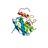
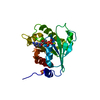
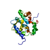
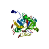
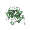
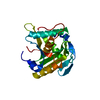
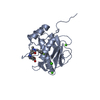
Crystal structure of human alpha1-acid glycoprotein
Components
Alpha-1-acid glycoprotein 1
Keywords
SIGNALING PROTEIN / PLASMA PROTEIN / GLYCOPROTEIN / POLYMORPHISM / ACUTE PHASE PROTEIN / SECRETED / PYRROLIDONE CARBOXYLIC ACID / LIPOCALIN
Function / homology
Function and homology information
positive regulation of interleukin-1 production / regulation of immune system process / negative regulation of interleukin-6 production / negative regulation of tumor necrosis factor production / platelet alpha granule lumen / acute-phase response / positive regulation of interleukin-1 beta production / specific granule lumen / positive regulation of tumor necrosis factor production / tertiary granule lumen ...positive regulation of interleukin-1 production / regulation of immune system process / negative regulation of interleukin-6 production / negative regulation of tumor necrosis factor production / platelet alpha granule lumen / acute-phase response / positive regulation of interleukin-1 beta production / specific granule lumen / positive regulation of tumor necrosis factor production / tertiary granule lumen / Platelet degranulation / blood microparticle / inflammatory response / Neutrophil degranulation / : / extracellular exosome / extracellular region Similarity search - Function
In the structure databanks used in Yorodumi, some data are registered as the other names, "COVID-19 virus" and "2019-nCoV". Here are the details of the virus and the list of structure data.
Jan 31, 2019. EMDB accession codes are about to change! (news from PDBe EMDB page)
EMDB accession codes are about to change! (news from PDBe EMDB page)
The allocation of 4 digits for EMDB accession codes will soon come to an end. Whilst these codes will remain in use, new EMDB accession codes will include an additional digit and will expand incrementally as the available range of codes is exhausted. The current 4-digit format prefixed with “EMD-” (i.e. EMD-XXXX) will advance to a 5-digit format (i.e. EMD-XXXXX), and so on. It is currently estimated that the 4-digit codes will be depleted around Spring 2019, at which point the 5-digit format will come into force.
The EM Navigator/Yorodumi systems omit the EMD- prefix.
Related info.:Q: What is EMD? / ID/Accession-code notation in Yorodumi/EM Navigator
Yorodumi is a browser for structure data from EMDB, PDB, SASBDB, etc.
This page is also the successor to EM Navigator detail page, and also detail information page/front-end page for Omokage search.
The word "yorodu" (or yorozu) is an old Japanese word meaning "ten thousand". "mi" (miru) is to see.
Related info.:EMDB / PDB / SASBDB / Comparison of 3 databanks / Yorodumi Search / Aug 31, 2016. New EM Navigator & Yorodumi / Yorodumi Papers / Jmol/JSmol / Function and homology information / Changes in new EM Navigator and Yorodumi
 Movie
Movie Controller


 Open data
 Basic information
 Components
 Keywords
 Function and homology information
 Homo sapiens (human)
 X-RAY DIFFRACTION /
  Authors
 Citation
 Structure visualization
 Downloads & links
 Other downloads
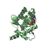


 PDBj
PDBj

 Assembly
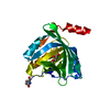


 Mass: 134.130 Da / Num. of mol.: 1 / Source method: obtained synthetically / Formula: C5H10O4
 Mass: 35.453 Da / Num. of mol.: 1 / Source method: obtained synthetically / Formula: Cl
 Mass: 18.015 Da / Num. of mol.: 142 / Source method: isolated from a natural source / Formula: H2O
 Sample preparation

 Processing