 Movie
Movie Controller


+ Open data

- Basic information
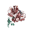
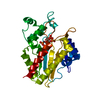
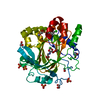
| Entry | Database: PDB / ID: 3kp6 | ||||||
|---|---|---|---|---|---|---|---|
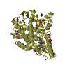
| Title | Staphylococcus epidermidis TcaR in complex with salicylate | ||||||
 Components Components | Transcriptional regulator TcaR | ||||||
 Keywords Keywords | TRANSCRIPTION REGULATOR / Multiple drug resistance / biofilm / transcription regulation / DNA binding / antibiotics / Transcription | ||||||
| Function / homology |  Function and homology information Function and homology informationresponse to stress / DNA-binding transcription factor activity / DNA binding Similarity search - Function | ||||||
| Biological species |  Staphylococcus epidermidis RP62A (bacteria) Staphylococcus epidermidis RP62A (bacteria) | ||||||
| Method |  X-RAY DIFFRACTION / SYNCHROTRON / MOLECULAR REPLACEMENT / Resolution: 2.45 Å X-RAY DIFFRACTION / SYNCHROTRON / MOLECULAR REPLACEMENT / Resolution: 2.45 Å | ||||||
 Authors Authors | Chang, Y.M. / Chen, C.K. / Yeh, Y.J. / Wang, A.H. | ||||||
 Citation Citation | Journal: Proc.Natl.Acad.Sci.USA / Year: 2010 Title: Structural study of TcaR and its complexes with multiple antibiotics from Staphylococcus epidermidis. Authors: Chang, Y.M. / Jeng, W.Y. / Ko, T.P. / Yeh, Y.J. / Chen, C.K. / Wang, A.H. | ||||||
| History |
|
- Structure visualization
| Structure viewer | Molecule: MolmilJmol/JSmol |
|---|
- Downloads & links
-Download
| PDBx/mmCIF format | 3kp6.cif.gz | 71.6 KB | Display | PDBx/mmCIF format |
|---|---|---|---|---|
| PDB format | pdb3kp6.ent.gz | 54.1 KB | Display | PDB format |
| PDBx/mmJSON format | 3kp6.json.gz | Tree view | PDBx/mmJSON format | |
| Others |  Other downloads Other downloads |
-Validation report
| Summary document | 3kp6_validation.pdf.gz | 471.9 KB | Display | wwPDB validaton report |
|---|---|---|---|---|
| Full document | 3kp6_full_validation.pdf.gz | 486.8 KB | Display | |
| Data in XML | 3kp6_validation.xml.gz | 14.9 KB | Display | |
| Data in CIF | 3kp6_validation.cif.gz | 19.7 KB | Display | |
| Arichive directory | https://data.pdbj.org/pub/pdb/validation_reports/kp/3kp6ftp://data.pdbj.org/pub/pdb/validation_reports/kp/3kp6 | HTTPS FTP |
-Related structure data
| Related structure data |  3kp2C  3kp3C  3kp4C  3kp5C  3kp7SC C: citing same article ( S: Starting model for refinement |
|---|---|
| Similar structure data |


-Links
 PDBj
PDBj


- Assembly
| Deposited unit | 
| ||||||||
|---|---|---|---|---|---|---|---|---|---|
| 1 |
| ||||||||
| Unit cell |
|
-Components
| #1: Protein | Mass: 17378.195 Da / Num. of mol.: 2 Source method: isolated from a genetically manipulated source Source: (gene. exp.) Staphylococcus epidermidis RP62A (bacteria)Strain: ATCC 35984 / Gene: SERP1949, SE_1937, tcaR / Plasmid: pET 21a / Production host: #2: Chemical | ChemComp-SAL / 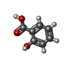  Mass: 138.121 Da / Num. of mol.: 8 / Source method: obtained synthetically / Formula: C7H6O3 Mass: 138.121 Da / Num. of mol.: 8 / Source method: obtained synthetically / Formula: C7H6O3#3: Chemical | ChemComp-CAC / |   Mass: 136.989 Da / Num. of mol.: 1 / Source method: obtained synthetically / Formula: C2H6AsO2 Mass: 136.989 Da / Num. of mol.: 1 / Source method: obtained synthetically / Formula: C2H6AsO2#4: Water | ChemComp-HOH / |  Mass: 18.015 Da / Num. of mol.: 80 / Source method: isolated from a natural source / Formula: H2O Mass: 18.015 Da / Num. of mol.: 80 / Source method: isolated from a natural source / Formula: H2O |
|---|
-Experimental details
-Experiment
| Experiment | Method: X-RAY DIFFRACTION / Number of used crystals: 1 |
|---|
- Sample preparation
| Crystal | Density Matthews: 2.4 Å3/Da / Density % sol: 48.67 % |
|---|---|
| Crystal grow | Temperature: 298 K / Method: vapor diffusion, hanging drop / pH: 7.5 Details: 0.1M Na-Hepes, pH 7.5, 8-14% PEG 4000, 10-13% 2-propanol precipitant, VAPOR DIFFUSION, HANGING DROP, temperature 298K |
-Data collection
| Diffraction | Mean temperature: 100 K |
|---|---|
| Diffraction source | Source: SYNCHROTRON / Site: NSRRC  / Beamline: BL13B1 / Wavelength: 1 Å / Beamline: BL13B1 / Wavelength: 1 Å |
| Detector | Type: ADSC QUANTUM 210 / Detector: CCD / Date: Jan 10, 2008 / Details: mirrors |
| Radiation | Monochromator: GRAPHITE / Protocol: SINGLE WAVELENGTH / Monochromatic (M) / Laue (L): M / Scattering type: x-ray |
| Radiation wavelength | Wavelength: 1 Å / Relative weight: 1 |
| Reflection | Resolution: 2.45→30 Å / Num. all: 12267 / Num. obs: 12192 / % possible obs: 99.4 % / Observed criterion σ(F): 0 / Observed criterion σ(I): 0 / Redundancy: 13.9 % / Rmerge(I) obs: 0.073 / Net I/σ(I): 34.3 |
| Reflection shell | Resolution: 2.45→2.54 Å / Redundancy: 11 % / Rmerge(I) obs: 0.551 / Mean I/σ(I) obs: 2.1 / Num. unique all: 1126 / % possible all: 77.5 |
- Processing
| Software |
| |||||||||||||||||||||||||
|---|---|---|---|---|---|---|---|---|---|---|---|---|---|---|---|---|---|---|---|---|---|---|---|---|---|---|
| Refinement | Method to determine structure: MOLECULAR REPLACEMENT Starting model: 3KP7 Resolution: 2.45→30 Å / Isotropic thermal model: Isotropic / Cross valid method: THROUGHOUT / σ(F): 0 / Stereochemistry target values: Engh & Huber
| |||||||||||||||||||||||||
| Displacement parameters | Biso mean: 60.82 Å2 | |||||||||||||||||||||||||
| Refinement step | Cycle: LAST / Resolution: 2.45→30 Å
| |||||||||||||||||||||||||
| Refine LS restraints |
| |||||||||||||||||||||||||
| LS refinement shell | Resolution: 2.45→2.54 Å
|
