 Movie
Movie Controller

[English] 日本語
 Yorodumi
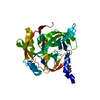
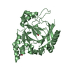
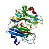
- PDB-3kn1: Crystal Structure of Golgi Phosphoprotein 3 N-term Truncation Variant -
+ Open data

- Basic information
| Entry | Database: PDB / ID: 3kn1 | ||||||
|---|---|---|---|---|---|---|---|
| Title | Crystal Structure of Golgi Phosphoprotein 3 N-term Truncation Variant | ||||||
 Components Components | Golgi phosphoprotein 3 | ||||||
 Keywords Keywords | PROTEIN BINDING / beta hairpin / phosphoinositide binding domain / Cell membrane / Cytoplasm / Endosome / Golgi apparatus / Membrane / Mitochondrion / Phosphoprotein | ||||||
| Function / homology |  Function and homology information Function and homology informationasymmetric Golgi ribbon formation / protein targeting to Golgi apparatus / Golgi vesicle budding / cargo adaptor activity / cellular response to rapamycin / Golgi cisterna / protein retention in Golgi apparatus / Golgi ribbon formation / cell adhesion molecule production / leukocyte tethering or rolling ...asymmetric Golgi ribbon formation / protein targeting to Golgi apparatus / Golgi vesicle budding / cargo adaptor activity / cellular response to rapamycin / Golgi cisterna / protein retention in Golgi apparatus / Golgi ribbon formation / cell adhesion molecule production / leukocyte tethering or rolling / glycoprotein biosynthetic process / Golgi to plasma membrane protein transport / retrograde vesicle-mediated transport, Golgi to endoplasmic reticulum / regulation of mitochondrion organization / phosphatidylinositol-4-phosphate binding / lamellipodium assembly / Golgi cisterna membrane / Golgi organization / protein secretion / positive regulation of TOR signaling / positive regulation of protein secretion / trans-Golgi network / mitochondrial intermembrane space / cell migration / gene expression / endosome / Golgi membrane / negative regulation of apoptotic process / enzyme binding / Golgi apparatus / mitochondrion / plasma membrane / cytosol Similarity search - Function | ||||||
| Biological species |  Homo sapiens (human) Homo sapiens (human) | ||||||
| Method |  X-RAY DIFFRACTION / SYNCHROTRON / MOLECULAR REPLACEMENT / molecular replacement / Resolution: 2.9 Å X-RAY DIFFRACTION / SYNCHROTRON / MOLECULAR REPLACEMENT / molecular replacement / Resolution: 2.9 Å | ||||||
 Authors Authors | Schmitz, K.R. / Bessman, N.J. / Setty, T.G. / Ferguson, K.M. | ||||||
 Citation Citation | Journal: J.Cell Biol. / Year: 2009 Title: PtdIns4P recognition by Vps74/GOLPH3 links PtdIns 4-kinase signaling to retrograde Golgi trafficking. Authors: Wood, C.S. / Schmitz, K.R. / Bessman, N.J. / Setty, T.G. / Ferguson, K.M. / Burd, C.G. | ||||||
| History |
|
- Structure visualization
| Structure viewer | Molecule: MolmilJmol/JSmol |
|---|
- Downloads & links
-Download
| PDBx/mmCIF format | 3kn1.cif.gz | 63.4 KB | Display | PDBx/mmCIF format |
|---|---|---|---|---|
| PDB format | pdb3kn1.ent.gz | 46.5 KB | Display | PDB format |
| PDBx/mmJSON format | 3kn1.json.gz | Tree view | PDBx/mmJSON format | |
| Others |  Other downloads Other downloads |
-Validation report
| Arichive directory | https://data.pdbj.org/pub/pdb/validation_reports/kn/3kn1ftp://data.pdbj.org/pub/pdb/validation_reports/kn/3kn1 | HTTPS FTP |
|---|
-Related structure data
| Related structure data |  2zihS S: Starting model for refinement |
|---|---|
| Similar structure data |


-Links
 PDBj
PDBj- Assembly
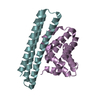
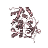
| Deposited unit | 
| ||||||||
|---|---|---|---|---|---|---|---|---|---|
| 1 |
| ||||||||
| 2 | x 24
| ||||||||
| Unit cell |
| ||||||||
| Details | authors state that the software-determined 24-mer involves interactions stabilized by non-physiological inter-copy disulfide bonds between symmetry-related copies of chain A. |
-Components
| #1: Protein | Mass: 28792.574 Da / Num. of mol.: 1 / Fragment: UNP residues 52-298 Source method: isolated from a genetically manipulated source Source: (gene. exp.) Homo sapiens (human) / Gene: GOLPH3, GPP34 / Plasmid: pET-21a / Production host:  | ||||
|---|---|---|---|---|---|
| #2: Chemical | ChemComp-SO4 /   Mass: 96.063 Da / Num. of mol.: 6 / Source method: obtained synthetically / Formula: SO4 Mass: 96.063 Da / Num. of mol.: 6 / Source method: obtained synthetically / Formula: SO4#3: Water | ChemComp-HOH / |  Mass: 18.015 Da / Num. of mol.: 9 / Source method: isolated from a natural source / Formula: H2O Mass: 18.015 Da / Num. of mol.: 9 / Source method: isolated from a natural source / Formula: H2OHas protein modification | Y | |
-Experimental details
-Experiment
| Experiment | Method: X-RAY DIFFRACTION / Number of used crystals: 1 |
|---|
- Sample preparation
| Crystal | Density Matthews: 3.84 Å3/Da / Density % sol: 67.94 % |
|---|---|
| Crystal grow | Temperature: 295 K / Method: vapor diffusion, hanging drop / pH: 6 Details: 0.9 M (NH4)2SO4, pH 6.0, VAPOR DIFFUSION, HANGING DROP, temperature 295K |
-Data collection
| Diffraction | Mean temperature: 100 K |
|---|---|
| Diffraction source | Source: SYNCHROTRON / Site: APS  / Beamline: 23-ID-B / Wavelength: 0.97951 Å / Beamline: 23-ID-B / Wavelength: 0.97951 Å |
| Detector | Type: MARMOSAIC 300 mm CCD / Detector: CCD / Date: Jul 16, 2008 |
| Radiation | Protocol: SINGLE WAVELENGTH / Monochromatic (M) / Laue (L): M / Scattering type: x-ray |
| Radiation wavelength | Wavelength: 0.97951 Å / Relative weight: 1 |
| Reflection | Resolution: 2.9→50 Å / Num. obs: 10180 / % possible obs: 98.3 % / Observed criterion σ(I): 2 / Redundancy: 3.6 % / Rmerge(I) obs: 0.133 / Χ2: 1.67 / Net I/σ(I): 7.8 |
| Reflection shell | Resolution: 2.9→3 Å / Redundancy: 3.6 % / Rmerge(I) obs: 0.586 / Mean I/σ(I) obs: 2.4 / Num. unique all: 994 / Χ2: 1.393 / % possible all: 99.6 |
-Phasing
| Phasing | Method: molecular replacement | |||||||||
|---|---|---|---|---|---|---|---|---|---|---|
| Phasing MR | Model details: Phaser MODE: MR_AUTO
|
- Processing
| Software |
| |||||||||||||||||||||||||||||||||||||||||||||||||||||||||||||||||
|---|---|---|---|---|---|---|---|---|---|---|---|---|---|---|---|---|---|---|---|---|---|---|---|---|---|---|---|---|---|---|---|---|---|---|---|---|---|---|---|---|---|---|---|---|---|---|---|---|---|---|---|---|---|---|---|---|---|---|---|---|---|---|---|---|---|---|
| Refinement | Method to determine structure: MOLECULAR REPLACEMENT Starting model: PDB entry 2ZIH Resolution: 2.9→50 Å / Cor.coef. Fo:Fc: 0.918 / Cor.coef. Fo:Fc free: 0.894 / WRfactor Rfree: 0.246 / WRfactor Rwork: 0.196 / Occupancy max: 1 / Occupancy min: 0.5 / FOM work R set: 0.816 / SU R Cruickshank DPI: 0.637 / SU Rfree: 0.354 / Cross valid method: THROUGHOUT / σ(F): 0 / ESU R: 0.637 / ESU R Free: 0.354 / Stereochemistry target values: MAXIMUM LIKELIHOOD Details: HYDROGENS HAVE BEEN ADDED IN THE RIDING POSITIONS U VALUES : REFINED INDIVIDUALLY
| |||||||||||||||||||||||||||||||||||||||||||||||||||||||||||||||||
| Solvent computation | Ion probe radii: 0.8 Å / Shrinkage radii: 0.8 Å / VDW probe radii: 1.4 Å / Solvent model: MASK | |||||||||||||||||||||||||||||||||||||||||||||||||||||||||||||||||
| Displacement parameters | Biso max: 109.27 Å2 / Biso mean: 38.227 Å2 / Biso min: 11.27 Å2 | |||||||||||||||||||||||||||||||||||||||||||||||||||||||||||||||||
| Refinement step | Cycle: LAST / Resolution: 2.9→50 Å
| |||||||||||||||||||||||||||||||||||||||||||||||||||||||||||||||||
| Refine LS restraints |
| |||||||||||||||||||||||||||||||||||||||||||||||||||||||||||||||||
| LS refinement shell | Resolution: 2.9→2.975 Å / Total num. of bins used: 20
|
