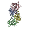
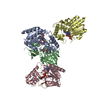
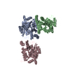
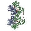
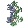
A: Vacuolar protein sorting-associated protein 74 B: Vacuolar protein sorting-associated protein 74 C: Vacuolar protein sorting-associated protein 74 D: Vacuolar protein sorting-associated protein 74
Redundancy: 10.2 % / Av σ(I) over netI: 6.9 / Number: 254363 / Rmerge(I) obs: 0.14 / Χ2: 1.76 / D res high: 3.4 Å / D res low: 50 Å / Num. obs: 24850 / % possible obs: 99
Diffraction reflection shell
Highest resolution (Å)
Lowest resolution (Å)
% possible obs (%)
ID
Rmerge(I) obs
Chi squared
Redundancy
7.32
50
99.3
1
0.065
3.956
10
5.81
7.32
99.6
1
0.125
2.322
10.7
5.08
5.81
99.7
1
0.14
1.796
10.9
4.61
5.08
99.6
1
0.128
1.698
11
4.28
4.61
99.5
1
0.134
1.574
11.1
4.03
4.28
99.5
1
0.164
1.333
11.1
3.83
4.03
99.4
1
0.218
1.203
11
3.66
3.83
99.2
1
0.282
1.098
10.5
3.52
3.66
98.6
1
0.378
1.097
9.2
3.4
3.52
95.4
1
0.456
1.031
6.6
Reflection
Resolution: 2.8→50 Å / Num. obs: 43558 / % possible obs: 97.4 % / Redundancy: 5.2 % / Biso Wilson estimate: 58.3 Å2 / Rmerge(I) obs: 0.093 / Χ2: 2.024 / Net I/σ(I): 11.3
Reflection shell
Resolution (Å)
Redundancy (%)
Rmerge(I) obs
Num. unique all
Χ2
% possible all
2.8-2.9
2.8
0.342
3658
1.282
83.8
2.9-3.02
3.5
0.307
4227
1.282
95.2
3.02-3.15
4.7
0.281
4343
1.254
99.3
3.15-3.32
5.6
0.225
4408
1.292
99.9
3.32-3.53
5.9
0.17
4419
1.612
99.8
3.53-3.8
5.9
0.123
4410
2.005
99.7
3.8-4.18
6
0.083
4445
2.166
99.8
4.18-4.79
6
0.067
4468
2.398
99.7
4.79-6.03
5.9
0.071
4537
2.734
99.6
6.03-50
5.6
0.05
4643
3.056
97.4
-
Phasing
Phasing
Method: MAD
-
Processing
Software
Name
Version
Classification
NB
DENZO
datareduction
SCALEPACK
datascaling
SHELX
phasing
REFMAC
refinement
PDB_EXTRACT
3.004
dataextraction
Blu-Ice
datacollection
HKL-2000
datareduction
HKL-2000
datascaling
SHELXD
phasing
Refinement
Method to determine structure: MAD / Resolution: 2.8→37.99 Å / Cor.coef. Fo:Fc: 0.928 / Cor.coef. Fo:Fc free: 0.876 / SU B: 15.113 / SU ML: 0.296 / Cross valid method: THROUGHOUT / σ(F): 0 / ESU R: 0.732 / ESU R Free: 0.395 / Stereochemistry target values: MAXIMUM LIKELIHOOD / Details: HYDROGENS HAVE BEEN ADDED IN THE RIDING POSITIONS
Rfactor
Num. reflection
% reflection
Selection details
Rfree
0.302
2198
5.1 %
RANDOM
Rwork
0.233
-
-
-
obs
0.237
43510
96.57 %
-
Solvent computation
Ion probe radii: 0.8 Å / Shrinkage radii: 0.8 Å / VDW probe radii: 1.2 Å / Solvent model: MASK
Displacement parameters
Biso mean: 45.287 Å2
Baniso -1
Baniso -2
Baniso -3
1-
1.94 Å2
0.97 Å2
0 Å2
2-
-
1.94 Å2
0 Å2
3-
-
-
-2.9 Å2
Refinement step
Cycle: LAST / Resolution: 2.8→37.99 Å
Protein
Nucleic acid
Ligand
Solvent
Total
Num. atoms
8536
0
0
5
8541
Refine LS restraints
Refine-ID
Type
Dev ideal
Dev ideal target
Number
X-RAY DIFFRACTION
r_bond_refined_d
0.013
0.022
8665
X-RAY DIFFRACTION
r_angle_refined_deg
1.64
1.976
11772
X-RAY DIFFRACTION
r_dihedral_angle_1_deg
7.317
5
1121
X-RAY DIFFRACTION
r_dihedral_angle_2_deg
38.039
23.906
361
X-RAY DIFFRACTION
r_dihedral_angle_3_deg
21.366
15
1451
X-RAY DIFFRACTION
r_dihedral_angle_4_deg
21.033
15
64
X-RAY DIFFRACTION
r_chiral_restr
0.126
0.2
1422
X-RAY DIFFRACTION
r_gen_planes_refined
0.005
0.02
6429
X-RAY DIFFRACTION
r_nbd_refined
0.244
0.2
4133
X-RAY DIFFRACTION
r_nbtor_refined
0.318
0.2
6104
X-RAY DIFFRACTION
r_xyhbond_nbd_refined
0.17
0.2
258
X-RAY DIFFRACTION
r_symmetry_vdw_refined
0.186
0.2
29
X-RAY DIFFRACTION
r_symmetry_hbond_refined
0.166
0.2
3
X-RAY DIFFRACTION
r_mcbond_it
0.617
1.5
5676
X-RAY DIFFRACTION
r_mcangle_it
1.137
2
8916
X-RAY DIFFRACTION
r_scbond_it
1.579
3
3296
X-RAY DIFFRACTION
r_scangle_it
2.646
4.5
2856
LS refinement shell
Resolution: 2.8→2.861 Å / Total num. of bins used: 20
Rfactor
Num. reflection
% reflection
Rfree
0.422
116
-
Rwork
0.329
1820
-
all
-
1936
-
obs
-
-
69.32 %
+
About Yorodumi
-
News
-
Feb 9, 2022. New format data for meta-information of EMDB entries
New format data for meta-information of EMDB entries
Version 3 of the EMDB header file is now the official format.
The previous official version 1.9 will be removed from the archive.
In the structure databanks used in Yorodumi, some data are registered as the other names, "COVID-19 virus" and "2019-nCoV". Here are the details of the virus and the list of structure data.
Jan 31, 2019. EMDB accession codes are about to change! (news from PDBe EMDB page)
EMDB accession codes are about to change! (news from PDBe EMDB page)
The allocation of 4 digits for EMDB accession codes will soon come to an end. Whilst these codes will remain in use, new EMDB accession codes will include an additional digit and will expand incrementally as the available range of codes is exhausted. The current 4-digit format prefixed with “EMD-” (i.e. EMD-XXXX) will advance to a 5-digit format (i.e. EMD-XXXXX), and so on. It is currently estimated that the 4-digit codes will be depleted around Spring 2019, at which point the 5-digit format will come into force.
The EM Navigator/Yorodumi systems omit the EMD- prefix.
Related info.:Q: What is EMD? / ID/Accession-code notation in Yorodumi/EM Navigator
Yorodumi is a browser for structure data from EMDB, PDB, SASBDB, etc.
This page is also the successor to EM Navigator detail page, and also detail information page/front-end page for Omokage search.
The word "yorodu" (or yorozu) is an old Japanese word meaning "ten thousand". "mi" (miru) is to see.
Related info.:EMDB / PDB / SASBDB / Comparison of 3 databanks / Yorodumi Search / Aug 31, 2016. New EM Navigator & Yorodumi / Yorodumi Papers / Jmol/JSmol / Function and homology information / Changes in new EM Navigator and Yorodumi
 Movie
Movie Controller


 Open data
 Basic information
 Components
 Keywords
 Function and homology information
 X-RAY DIFFRACTION /
  Authors
 Citation
 Structure visualization
 Downloads & links
 Other downloads


 PDBj
PDBj Assembly
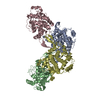

 Mass: 18.015 Da / Num. of mol.: 5 / Source method: isolated from a natural source / Formula: H2O
 Sample preparation
 Processing