 Movie
Movie Controller

[English] 日本語
 Yorodumi
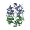
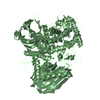
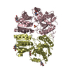
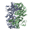
- PDB-2wjx: Crystal structure of the human ionotropic glutamate receptor GluR... -
+ Open data

- Basic information
| Entry | Database: PDB / ID: 2wjx | ||||||
|---|---|---|---|---|---|---|---|
| Title | Crystal structure of the human ionotropic glutamate receptor GluR2 ATD region at 4.1 A resolution | ||||||
 Components Components | GLUTAMATE RECEPTOR 2 | ||||||
 Keywords Keywords | TRANSPORT PROTEIN / POSTSYNAPTIC CELL MEMBRANE / GLUR2 / SYNAPSE / MEMBRANE / RECEPTOR / PALMITATE / SYNAPTIC PLASTICITY / ALTERNATIVE SPLICING / ION TRANSPORT / CELL JUNCTION / CELL MEMBRANE / TRANSMEMBRANE / PHOSPHOPROTEIN / GLUTAMATE RECEPTORS / POLYMORPHISM / GLYCOPROTEIN / IONIC CHANNEL / TRANSPORT / ION CHANNEL / RNA EDITING / LIPOPROTEIN | ||||||
| Function / homology |  Function and homology information Function and homology informationActivation of AMPA receptors / Trafficking of GluR2-containing AMPA receptors / postsynaptic endocytic zone / ligand-gated monoatomic cation channel activity / AMPA glutamate receptor activity / Unblocking of NMDA receptors, glutamate binding and activation / AMPA glutamate receptor complex / Long-term potentiation / asymmetric synapse / MECP2 regulates neuronal receptors and channels ...Activation of AMPA receptors / Trafficking of GluR2-containing AMPA receptors / postsynaptic endocytic zone / ligand-gated monoatomic cation channel activity / AMPA glutamate receptor activity / Unblocking of NMDA receptors, glutamate binding and activation / AMPA glutamate receptor complex / Long-term potentiation / asymmetric synapse / MECP2 regulates neuronal receptors and channels / glutamate-gated receptor activity / excitatory synapse / ionotropic glutamate receptor signaling pathway / transmitter-gated monoatomic ion channel activity involved in regulation of postsynaptic membrane potential / synaptic transmission, glutamatergic / postsynaptic density membrane / modulation of chemical synaptic transmission / endocytic vesicle membrane / amyloid-beta binding / dendritic spine / postsynaptic density / postsynapse / external side of plasma membrane / neuronal cell body / dendrite / plasma membrane Similarity search - Function | ||||||
| Biological species |  HOMO SAPIENS (human) HOMO SAPIENS (human) | ||||||
| Method |  X-RAY DIFFRACTION / SYNCHROTRON / MOLECULAR REPLACEMENT / Resolution: 4.1 Å X-RAY DIFFRACTION / SYNCHROTRON / MOLECULAR REPLACEMENT / Resolution: 4.1 Å | ||||||
 Authors Authors | Clayton, A. / Siebold, C. / Gilbert, R.J.C. / Sutton, G.C. / Harlos, K. / McIlhinney, R.A.J. / Jones, E.Y. / Aricescu, A.R. | ||||||
 Citation Citation | Journal: J.Mol.Biol. / Year: 2009 Title: Crystal Structure of the Glur2 Amino-Terminal Domain Provides Insights Into the Architecture and Assembly of Ionotropic Glutamate Receptors. Authors: Clayton, A. / Siebold, C. / Gilbert, R.J.C. / Sutton, G.C. / Harlos, K. / Mcilhinney, R.A.J. / Jones, E.Y. / Aricescu, A.R. | ||||||
| History |
|
- Structure visualization
| Structure viewer | Molecule: MolmilJmol/JSmol |
|---|
- Downloads & links
-Download
| PDBx/mmCIF format | 2wjx.cif.gz | 456.5 KB | Display | PDBx/mmCIF format |
|---|---|---|---|---|
| PDB format | pdb2wjx.ent.gz | 385.2 KB | Display | PDB format |
| PDBx/mmJSON format | 2wjx.json.gz | Tree view | PDBx/mmJSON format | |
| Others |  Other downloads Other downloads |
-Validation report
| Arichive directory | https://data.pdbj.org/pub/pdb/validation_reports/wj/2wjxftp://data.pdbj.org/pub/pdb/validation_reports/wj/2wjx | HTTPS FTP |
|---|
-Related structure data


-Links
 PDBj
PDBj


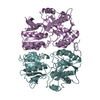
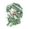
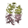
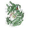
- Assembly
| Deposited unit | 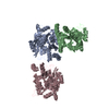
| |||||||||||||||||||||||||||||||||||||||||||||||||||||||||
|---|---|---|---|---|---|---|---|---|---|---|---|---|---|---|---|---|---|---|---|---|---|---|---|---|---|---|---|---|---|---|---|---|---|---|---|---|---|---|---|---|---|---|---|---|---|---|---|---|---|---|---|---|---|---|---|---|---|---|
| 1 | 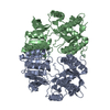
| |||||||||||||||||||||||||||||||||||||||||||||||||||||||||
| 2 | 
| |||||||||||||||||||||||||||||||||||||||||||||||||||||||||
| Unit cell |
| |||||||||||||||||||||||||||||||||||||||||||||||||||||||||
| Noncrystallographic symmetry (NCS) | NCS domain:
NCS domain segments:
NCS ensembles :
NCS oper:
|
-Components
| #1: Protein | Mass: 43775.328 Da / Num. of mol.: 3 / Fragment: AMINO-TERMINAL DOMAIN, RESIDUES 25-412 Source method: isolated from a genetically manipulated source Source: (gene. exp.) HOMO SAPIENS (human) / Plasmid: PHLSEC / Cell line (production host): HEK 293S GNTI- CELLS / Production host: HOMO SAPIENS (human) / References: UniProt: P42262Has protein modification | Y | |
|---|
-Experimental details
-Experiment
| Experiment | Method: X-RAY DIFFRACTION / Number of used crystals: 1 |
|---|
- Sample preparation
| Crystal | Density Matthews: 3.58 Å3/Da / Density % sol: 66 % / Description: NONE |
|---|---|
| Crystal grow | Details: 0.2M BIS-TRIS PROPANE, PH 7.5 0.2M POTASSIUM/SODIUM TARTRATE 20% (W/V) PEG3350 |
-Data collection
| Diffraction | Mean temperature: 100 K |
|---|---|
| Diffraction source | Source: SYNCHROTRON / Site: ESRF  / Beamline: ID29 / Wavelength: 0.9762 / Beamline: ID29 / Wavelength: 0.9762 |
| Detector | Type: ADSC CCD / Detector: CCD / Date: Oct 4, 2008 |
| Radiation | Protocol: SINGLE WAVELENGTH / Monochromatic (M) / Laue (L): M / Scattering type: x-ray |
| Radiation wavelength | Wavelength: 0.9762 Å / Relative weight: 1 |
| Reflection | Resolution: 4.1→50 Å / Num. obs: 15949 / % possible obs: 99.6 % / Observed criterion σ(I): 0 / Redundancy: 28.5 % / Biso Wilson estimate: 149.1 Å2 / Rmerge(I) obs: 0.12 / Net I/σ(I): 23.3 |
| Reflection shell | Resolution: 4.1→4.2 Å / Redundancy: 29.2 % / Rmerge(I) obs: 0.84 / Mean I/σ(I) obs: 5.7 / % possible all: 100 |
- Processing
| Software |
| |||||||||||||||||||||||||||||||||||||||||||||||||||||||||||||||||||||||||||||||||||||||||||||||||||||||||||||||||||||||||||||||||||||||||||||||||||||||||||||||||||||||||||||||
|---|---|---|---|---|---|---|---|---|---|---|---|---|---|---|---|---|---|---|---|---|---|---|---|---|---|---|---|---|---|---|---|---|---|---|---|---|---|---|---|---|---|---|---|---|---|---|---|---|---|---|---|---|---|---|---|---|---|---|---|---|---|---|---|---|---|---|---|---|---|---|---|---|---|---|---|---|---|---|---|---|---|---|---|---|---|---|---|---|---|---|---|---|---|---|---|---|---|---|---|---|---|---|---|---|---|---|---|---|---|---|---|---|---|---|---|---|---|---|---|---|---|---|---|---|---|---|---|---|---|---|---|---|---|---|---|---|---|---|---|---|---|---|---|---|---|---|---|---|---|---|---|---|---|---|---|---|---|---|---|---|---|---|---|---|---|---|---|---|---|---|---|---|---|---|---|---|
| Refinement | Method to determine structure: MOLECULAR REPLACEMENT / Resolution: 4.1→48.379 Å / SU ML: 0.46 / σ(F): 0.23 / Phase error: 34.74 / Stereochemistry target values: ML
| |||||||||||||||||||||||||||||||||||||||||||||||||||||||||||||||||||||||||||||||||||||||||||||||||||||||||||||||||||||||||||||||||||||||||||||||||||||||||||||||||||||||||||||||
| Solvent computation | Shrinkage radii: 0.9 Å / VDW probe radii: 1.11 Å / Solvent model: FLAT BULK SOLVENT MODEL / Bsol: 99 Å2 / ksol: 0.311 e/Å3 | |||||||||||||||||||||||||||||||||||||||||||||||||||||||||||||||||||||||||||||||||||||||||||||||||||||||||||||||||||||||||||||||||||||||||||||||||||||||||||||||||||||||||||||||
| Displacement parameters |
| |||||||||||||||||||||||||||||||||||||||||||||||||||||||||||||||||||||||||||||||||||||||||||||||||||||||||||||||||||||||||||||||||||||||||||||||||||||||||||||||||||||||||||||||
| Refinement step | Cycle: LAST / Resolution: 4.1→48.379 Å
| |||||||||||||||||||||||||||||||||||||||||||||||||||||||||||||||||||||||||||||||||||||||||||||||||||||||||||||||||||||||||||||||||||||||||||||||||||||||||||||||||||||||||||||||
| Refine LS restraints |
| |||||||||||||||||||||||||||||||||||||||||||||||||||||||||||||||||||||||||||||||||||||||||||||||||||||||||||||||||||||||||||||||||||||||||||||||||||||||||||||||||||||||||||||||
| Refine LS restraints NCS |
| |||||||||||||||||||||||||||||||||||||||||||||||||||||||||||||||||||||||||||||||||||||||||||||||||||||||||||||||||||||||||||||||||||||||||||||||||||||||||||||||||||||||||||||||
| LS refinement shell |
| |||||||||||||||||||||||||||||||||||||||||||||||||||||||||||||||||||||||||||||||||||||||||||||||||||||||||||||||||||||||||||||||||||||||||||||||||||||||||||||||||||||||||||||||
| Refinement TLS params. | Method: refined / Refine-ID: X-RAY DIFFRACTION
| |||||||||||||||||||||||||||||||||||||||||||||||||||||||||||||||||||||||||||||||||||||||||||||||||||||||||||||||||||||||||||||||||||||||||||||||||||||||||||||||||||||||||||||||
| Refinement TLS group |
|
