 Movie
Movie Controller

[English] 日本語
 Yorodumi
- PDB-3ke7: Crystal structure of putative ketosteroid isomerase (YP_001303366... -
+ Open data

- Basic information
| Entry | Database: PDB / ID: 3ke7 | ||||||
|---|---|---|---|---|---|---|---|
| Title | Crystal structure of putative ketosteroid isomerase (YP_001303366.1) from Parabacteroides distasonis ATCC 8503 at 1.45 A resolution | ||||||
 Components Components | Putative ketosteroid isomerase | ||||||
 Keywords Keywords | ISOMERASE / putative ketosteroid isomerase / Structural Genomics / Joint Center for Structural Genomics / JCSG / Protein Structure Initiative / PSI-2 | ||||||
| Function / homology | SnoaL-like domain / SnoaL-like domain / Nuclear Transport Factor 2; Chain: A, - #50 / NTF2-like domain superfamily / Nuclear Transport Factor 2; Chain: A, / Roll / Alpha Beta / DI(HYDROXYETHYL)ETHER / SnoaL-like domain-containing protein Function and homology information Function and homology information | ||||||
| Biological species |  Parabacteroides distasonis ATCC 8503 (bacteria) Parabacteroides distasonis ATCC 8503 (bacteria) | ||||||
| Method |  X-RAY DIFFRACTION / SYNCHROTRON / MAD / Resolution: 1.45 Å X-RAY DIFFRACTION / SYNCHROTRON / MAD / Resolution: 1.45 Å | ||||||
 Authors Authors | Joint Center for Structural Genomics (JCSG) | ||||||
 Citation Citation | Journal: To be published Title: Crystal structure of putative ketosteroid isomerase (YP_001303366.1) from Parabacteroides distasonis ATCC 8503 at 1.45 A resolution Authors: Joint Center for Structural Genomics (JCSG) | ||||||
| History |
|
- Structure visualization
| Structure viewer | Molecule: MolmilJmol/JSmol |
|---|
- Downloads & links
-Download
| PDBx/mmCIF format | 3ke7.cif.gz | 79.6 KB | Display | PDBx/mmCIF format |
|---|---|---|---|---|
| PDB format | pdb3ke7.ent.gz | 59.6 KB | Display | PDB format |
| PDBx/mmJSON format | 3ke7.json.gz | Tree view | PDBx/mmJSON format | |
| Others |  Other downloads Other downloads |
-Validation report
| Arichive directory | https://data.pdbj.org/pub/pdb/validation_reports/ke/3ke7ftp://data.pdbj.org/pub/pdb/validation_reports/ke/3ke7 | HTTPS FTP |
|---|
-Related structure data
| Similar structure data | |
|---|---|
| Other databases |


-Links
 PDBj
PDBj
- Assembly
| Deposited unit | 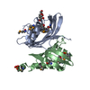
| ||||||||
|---|---|---|---|---|---|---|---|---|---|
| 1 |
| ||||||||
| Unit cell |
| ||||||||
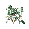
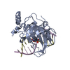
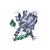
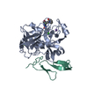
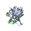
| Details | CRYSTAL PACKING AND ANALYTICAL SIZE EXCLUSION CHROMATOGRAPHY ANALYSES SUPPORT THE ASSIGNMENT OF A DIMER AS A SIGNIFICANT OLIGOMERIZATION STATE IN SOLUTION. |
-Components
-Protein , 1 types, 2 molecules AB
| #1: Protein | Mass: 15874.207 Da / Num. of mol.: 2 / Fragment: sequence database residues 22-154 Source method: isolated from a genetically manipulated source Source: (gene. exp.) Parabacteroides distasonis ATCC 8503 (bacteria)Strain: ATCC 8503 / DSM 20701 / NCTC 11152 / Gene: BDI_2009 / Plasmid: SpeedET / Production host: |
|---|
-Non-polymers , 5 types, 252 molecules 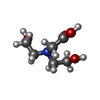


| #2: Chemical |  Mass: 163.172 Da / Num. of mol.: 2 / Source method: obtained synthetically / Formula: C6H13NO4 / Comment: pH buffer*YM Mass: 163.172 Da / Num. of mol.: 2 / Source method: obtained synthetically / Formula: C6H13NO4 / Comment: pH buffer*YM#3: Chemical | ChemComp-GOL / |  Mass: 92.094 Da / Num. of mol.: 1 / Source method: obtained synthetically / Formula: C3H8O3 Mass: 92.094 Da / Num. of mol.: 1 / Source method: obtained synthetically / Formula: C3H8O3#4: Chemical | ChemComp-PEG / |  Mass: 106.120 Da / Num. of mol.: 1 / Source method: obtained synthetically / Formula: C4H10O3 Mass: 106.120 Da / Num. of mol.: 1 / Source method: obtained synthetically / Formula: C4H10O3#5: Chemical | ChemComp-SO4 / |  Mass: 96.063 Da / Num. of mol.: 1 / Source method: obtained synthetically / Formula: SO4 Mass: 96.063 Da / Num. of mol.: 1 / Source method: obtained synthetically / Formula: SO4#6: Water | ChemComp-HOH / | Mass: 18.015 Da / Num. of mol.: 247 / Source method: isolated from a natural source / Formula: H2O |
|---|
-Details
| Has protein modification | Y |
|---|---|
| Sequence details | THIS CONSTRUCT (22-154) WAS EXPRESSED WITH THE PURIFICATION TAG MGSDKIHHHHHHENLYFQG. THE TAG WAS ...THIS CONSTRUCT (22-154) WAS EXPRESSED WITH THE PURIFICATI |
-Experimental details
-Experiment
| Experiment | Method: X-RAY DIFFRACTION / Number of used crystals: 1 |
|---|
- Sample preparation
| Crystal | Density Matthews: 1.83 Å3/Da / Density % sol: 32.74 % |
|---|---|
| Crystal grow | Temperature: 277 K / Method: vapor diffusion, sitting drop / pH: 9 Details: 2.4000M (NH4)2SO4, 0.1M Bicine pH 9.0, NANODROP, VAPOR DIFFUSION, SITTING DROP, temperature 277K |
-Data collection
| Diffraction | Mean temperature: 100 K | |||||||||||||||||||||||||||||||||||||||||||||||||||||||||||||||||||||||||||||||||||||||||||||||||||||||||||||||||||||||||||||||||||||||||||||||||||
|---|---|---|---|---|---|---|---|---|---|---|---|---|---|---|---|---|---|---|---|---|---|---|---|---|---|---|---|---|---|---|---|---|---|---|---|---|---|---|---|---|---|---|---|---|---|---|---|---|---|---|---|---|---|---|---|---|---|---|---|---|---|---|---|---|---|---|---|---|---|---|---|---|---|---|---|---|---|---|---|---|---|---|---|---|---|---|---|---|---|---|---|---|---|---|---|---|---|---|---|---|---|---|---|---|---|---|---|---|---|---|---|---|---|---|---|---|---|---|---|---|---|---|---|---|---|---|---|---|---|---|---|---|---|---|---|---|---|---|---|---|---|---|---|---|---|---|---|---|
| Diffraction source | Source: SYNCHROTRON / Site: SSRL  / Beamline: BL11-1 / Wavelength: 0.91837,0.97805,0.97862 / Beamline: BL11-1 / Wavelength: 0.91837,0.97805,0.97862 | |||||||||||||||||||||||||||||||||||||||||||||||||||||||||||||||||||||||||||||||||||||||||||||||||||||||||||||||||||||||||||||||||||||||||||||||||||
| Detector | Type: MARMOSAIC 325 mm CCD / Detector: CCD / Date: Apr 16, 2009 / Details: Flat mirror (vertical focusing) | |||||||||||||||||||||||||||||||||||||||||||||||||||||||||||||||||||||||||||||||||||||||||||||||||||||||||||||||||||||||||||||||||||||||||||||||||||
| Radiation | Monochromator: Single crystal Si(111) bent monochromator (horizontal focusing) Protocol: MAD / Monochromatic (M) / Laue (L): M / Scattering type: x-ray | |||||||||||||||||||||||||||||||||||||||||||||||||||||||||||||||||||||||||||||||||||||||||||||||||||||||||||||||||||||||||||||||||||||||||||||||||||
| Radiation wavelength |
| |||||||||||||||||||||||||||||||||||||||||||||||||||||||||||||||||||||||||||||||||||||||||||||||||||||||||||||||||||||||||||||||||||||||||||||||||||
| Reflection | Resolution: 1.45→27.077 Å / Num. obs: 39881 / % possible obs: 98.5 % / Redundancy: 2.2 % / Biso Wilson estimate: 13.881 Å2 / Rmerge(I) obs: 0.065 / Rsym value: 0.065 / Net I/σ(I): 9.8 | |||||||||||||||||||||||||||||||||||||||||||||||||||||||||||||||||||||||||||||||||||||||||||||||||||||||||||||||||||||||||||||||||||||||||||||||||||
| Reflection shell | Diffraction-ID: 1 / Redundancy: 2.2 %
|
-Phasing
| Phasing | Method: MAD |
|---|
- Processing
| Software |
| |||||||||||||||||||||||||||||||||||||||||||||||||||||||||||||||||||||||||||||||||||||
|---|---|---|---|---|---|---|---|---|---|---|---|---|---|---|---|---|---|---|---|---|---|---|---|---|---|---|---|---|---|---|---|---|---|---|---|---|---|---|---|---|---|---|---|---|---|---|---|---|---|---|---|---|---|---|---|---|---|---|---|---|---|---|---|---|---|---|---|---|---|---|---|---|---|---|---|---|---|---|---|---|---|---|---|---|---|---|
| Refinement | Method to determine structure: MAD / Resolution: 1.45→27.077 Å / Cor.coef. Fo:Fc: 0.971 / Cor.coef. Fo:Fc free: 0.961 / Occupancy max: 1 / Occupancy min: 0.15 / SU B: 2.513 / SU ML: 0.043 / TLS residual ADP flag: LIKELY RESIDUAL / Cross valid method: THROUGHOUT / σ(F): 0 / ESU R: 0.069 / ESU R Free: 0.07 Stereochemistry target values: MAXIMUM LIKELIHOOD WITH PHASES Details: 1. HYDROGENS HAVE BEEN ADDED IN THE RIDING POSITIONS. 2. ATOM RECORD CONTAINS RESIDUAL B FACTORS ONLY. 3. A MET-INHIBITION PROTOCOL WAS USED FOR SELENOMETHIONINE INCORPORATION DURING PROTEIN ...Details: 1. HYDROGENS HAVE BEEN ADDED IN THE RIDING POSITIONS. 2. ATOM RECORD CONTAINS RESIDUAL B FACTORS ONLY. 3. A MET-INHIBITION PROTOCOL WAS USED FOR SELENOMETHIONINE INCORPORATION DURING PROTEIN EXPRESSION. THE OCCUPANCY OF THE SE ATOMS IN THE MSE RESIDUES WAS REDUCED TO 0.75 TO ACCOUNT FOR THE REDUCED SCATTERING POWER DUE TO PARTIAL S-MET INCORPORATION. 4. BICINE (BCN) AND GLYCEROL (GOL) FROM THE CRYSTALLIZATION/ CRYOPROTECTANT SOLUTIONS HAVE BEEN MODELED INTO THE SOLVENT STRUCTURE. 5. THE ELECTRON DENSITY FOR RESIDUES B119-B122 IS POOR. 6. THE CONFORMATION OF THE N-TERMINAL RESIDUES PRECEDING ASN-34 DIFFER CONSIDERABLY BETWEEN THE TWO NCS-RELATED PROTOMERS.
| |||||||||||||||||||||||||||||||||||||||||||||||||||||||||||||||||||||||||||||||||||||
| Solvent computation | Ion probe radii: 0.8 Å / Shrinkage radii: 0.8 Å / VDW probe radii: 1.4 Å / Solvent model: MASK | |||||||||||||||||||||||||||||||||||||||||||||||||||||||||||||||||||||||||||||||||||||
| Displacement parameters | Biso max: 61.55 Å2 / Biso mean: 13.291 Å2 / Biso min: 2 Å2
| |||||||||||||||||||||||||||||||||||||||||||||||||||||||||||||||||||||||||||||||||||||
| Refinement step | Cycle: LAST / Resolution: 1.45→27.077 Å
| |||||||||||||||||||||||||||||||||||||||||||||||||||||||||||||||||||||||||||||||||||||
| Refine LS restraints |
| |||||||||||||||||||||||||||||||||||||||||||||||||||||||||||||||||||||||||||||||||||||
| LS refinement shell | Resolution: 1.45→1.488 Å / Total num. of bins used: 20
| |||||||||||||||||||||||||||||||||||||||||||||||||||||||||||||||||||||||||||||||||||||
| Refinement TLS params. | Method: refined / Refine-ID: X-RAY DIFFRACTION
| |||||||||||||||||||||||||||||||||||||||||||||||||||||||||||||||||||||||||||||||||||||
| Refinement TLS group |
|
