 Movie
Movie Controller

[English] 日本語
 Yorodumi
- PDB-3kcw: Crystal structure of Ganoderma fungal immunomodulatory protein, GMI -
+ Open data

- Basic information
| Entry | Database: PDB / ID: 3kcw | ||||||
|---|---|---|---|---|---|---|---|
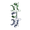
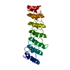
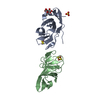
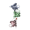
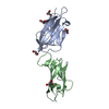
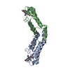
| Title | Crystal structure of Ganoderma fungal immunomodulatory protein, GMI | ||||||
 Components Components | immunomodulatory protein | ||||||
 Keywords Keywords | IMMUNE SYSTEM / FNIII | ||||||
| Function / homology |  Function and homology information Function and homology information | ||||||
| Biological species |  Ganoderma microsporum (fungus) Ganoderma microsporum (fungus) | ||||||
| Method |  X-RAY DIFFRACTION / SYNCHROTRON / MOLECULAR REPLACEMENT / Resolution: 2 Å X-RAY DIFFRACTION / SYNCHROTRON / MOLECULAR REPLACEMENT / Resolution: 2 Å | ||||||
 Authors Authors | Hsu, M.F. / Wang, A.H.J. / Yang, C.S. / Huang, C.T. / Hseu, R.S. / Lin, C.W. / Wu, M.Y. / Huang, C.S. / Fu, H.Y. | ||||||
 Citation Citation | Journal: To be Published Title: Single cysteine replacement at Leu6 increase the potent and thermostability in Ganoderma fungal immunomodulatory proteins Authors: Hsu, M.F. / Wang, A.H.J. / Yang, C.S. / Huang, C.T. / Hseu, R.S. / Lin, C.W. / Wu, M.Y. / Huang, C.S. / Fu, H.Y. | ||||||
| History |
|
- Structure visualization
| Structure viewer | Molecule: MolmilJmol/JSmol |
|---|
- Downloads & links
-Download
| PDBx/mmCIF format | 3kcw.cif.gz | 39 KB | Display | PDBx/mmCIF format |
|---|---|---|---|---|
| PDB format | pdb3kcw.ent.gz | 26.2 KB | Display | PDB format |
| PDBx/mmJSON format | 3kcw.json.gz | Tree view | PDBx/mmJSON format | |
| Others |  Other downloads Other downloads |
-Validation report
| Arichive directory | https://data.pdbj.org/pub/pdb/validation_reports/kc/3kcwftp://data.pdbj.org/pub/pdb/validation_reports/kc/3kcw | HTTPS FTP |
|---|
-Related structure data
| Related structure data | 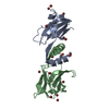 1osyS S: Starting model for refinement |
|---|---|
| Similar structure data |


-Links
 PDBj
PDBj- Assembly
| Deposited unit | 
| |||||||||||||||
|---|---|---|---|---|---|---|---|---|---|---|---|---|---|---|---|---|
| 1 | 
| |||||||||||||||
| Unit cell |
| |||||||||||||||
| Components on special symmetry positions |
|
-Components
| #1: Protein | Mass: 15185.717 Da / Num. of mol.: 1 Source method: isolated from a genetically manipulated source Source: (gene. exp.) Ganoderma microsporum (fungus) / Plasmid: pPICZaA / Production host: Pichia pastoris (fungus) / Strain (production host): KM71 / References: UniProt: E7FH75*PLUS |
|---|---|
| #2: Water | ChemComp-HOH /  Mass: 18.015 Da / Num. of mol.: 162 / Source method: isolated from a natural source / Formula: H2O Mass: 18.015 Da / Num. of mol.: 162 / Source method: isolated from a natural source / Formula: H2O |
| Sequence details | A SEQUENCE DATABASE REFERENCE FOR THIS PROTEIN DOES NOT CURRENTLY EXIST. |
-Experimental details
-Experiment
| Experiment | Method: X-RAY DIFFRACTION / Number of used crystals: 1 |
|---|
- Sample preparation
| Crystal | Density Matthews: 3.02 Å3/Da / Density % sol: 59.28 % |
|---|---|
| Crystal grow | Temperature: 298 K / Method: vapor diffusion, sitting drop / pH: 6.5 Details: 20% PEG 4000, 0.6M NaCl, 0.1M NaMES, pH 6.5, VAPOR DIFFUSION, SITTING DROP, temperature 298.0K |
-Data collection
| Diffraction | Mean temperature: 100 K |
|---|---|
| Diffraction source | Source: SYNCHROTRON / Site: NSRRC  / Beamline: BL13B1 / Wavelength: 1 Å / Beamline: BL13B1 / Wavelength: 1 Å |
| Detector | Type: ADSC QUANTUM 315 / Detector: CCD / Date: Jun 23, 2007 Details: Vertically Collimating Premirror, LN2-Cooled Fixed-Exit Double Crystal Si(111) Monochromator, Toroidal Focusing Mirror |
| Radiation | Monochromator: LN2-Cooled, Fixed-Exit Double Crystal Monochromator Protocol: SINGLE WAVELENGTH / Monochromatic (M) / Laue (L): M / Scattering type: x-ray |
| Radiation wavelength | Wavelength: 1 Å / Relative weight: 1 |
| Reflection | Resolution: 2→30 Å / Num. obs: 12868 / % possible obs: 100 % / Observed criterion σ(F): 0 / Observed criterion σ(I): 1 / Redundancy: 9.4 % / Biso Wilson estimate: 15.1 Å2 / Rmerge(I) obs: 0.09 / Rsym value: 0.251 |
| Reflection shell | Resolution: 2→2.07 Å / Redundancy: 9.3 % / Rmerge(I) obs: 0.737 / Mean I/σ(I) obs: 3.7 / Num. unique all: 1266 / % possible all: 100 |
- Processing
| Software |
| ||||||||||||||||||||||||||||||||||||
|---|---|---|---|---|---|---|---|---|---|---|---|---|---|---|---|---|---|---|---|---|---|---|---|---|---|---|---|---|---|---|---|---|---|---|---|---|---|
| Refinement | Method to determine structure: MOLECULAR REPLACEMENT Starting model: PDB enrty 1OSY Resolution: 2→25.85 Å / Rfactor Rfree error: 0.008 / Data cutoff high absF: 320890.2 / Data cutoff low absF: 0 / Isotropic thermal model: RESTRAINED / Cross valid method: THROUGHOUT / σ(F): 0 / Stereochemistry target values: Engh & Huber
| ||||||||||||||||||||||||||||||||||||
| Solvent computation | Solvent model: FLAT MODEL / Bsol: 53.5165 Å2 / ksol: 0.337558 e/Å3 | ||||||||||||||||||||||||||||||||||||
| Displacement parameters | Biso mean: 32 Å2
| ||||||||||||||||||||||||||||||||||||
| Refine analyze |
| ||||||||||||||||||||||||||||||||||||
| Refinement step | Cycle: LAST / Resolution: 2→25.85 Å
| ||||||||||||||||||||||||||||||||||||
| Refine LS restraints |
| ||||||||||||||||||||||||||||||||||||
| LS refinement shell | Resolution: 2→2.13 Å / Rfactor Rfree error: 0.025 / Total num. of bins used: 6
| ||||||||||||||||||||||||||||||||||||
| Xplor file |
|
