peptidyl-lysine hydroxylation to 5-hydroxy-L-lysine / histone H3R2 demethylase activity / peptidyl-lysine 5-dioxygenase activity / histone H4R3 demethylase activity / recognition of apoptotic cell / negative regulation of protein homooligomerization / oxidative RNA demethylation / oxidative RNA demethylase activity / protein demethylase activity / macrophage activation ...peptidyl-lysine hydroxylation to 5-hydroxy-L-lysine / histone H3R2 demethylase activity / peptidyl-lysine 5-dioxygenase activity / histone H4R3 demethylase activity / recognition of apoptotic cell / negative regulation of protein homooligomerization / oxidative RNA demethylation / oxidative RNA demethylase activity / protein demethylase activity / macrophage activation / regulation of mRNA splicing, via spliceosome / membraneless organelle assembly / transcription regulator activator activity / Oxidoreductases; Acting on paired donors, with incorporation or reduction of molecular oxygen; With 2-oxoglutarate as one donor, and incorporation of one atom of oxygen into each donor / sprouting angiogenesis / P-TEFb complex binding / histone demethylase activity / Protein hydroxylation / erythrocyte development / phagocytosis / lung development / RNA splicing / kidney development / HDMs demethylate histones / protein homooligomerization / mRNA processing / T cell differentiation in thymus / heart development / signaling receptor activity / retina development in camera-type eye / cell surface receptor signaling pathway / single-stranded RNA binding / chromatin remodeling / iron ion binding / ribonucleoprotein complex / nucleolus / positive regulation of DNA-templated transcription / positive regulation of transcription by RNA polymerase II / RNA binding / nucleoplasm / identical protein binding / nucleus / plasma membrane / cytosol / cytoplasm Similarity search - Function
Monooxygenase - #270 / : / Monooxygenase / Cupin / JmjC domain, hydroxylase / A domain family that is part of the cupin metalloenzyme superfamily. / JmjC domain / JmjC domain profile. / Jelly Rolls / Up-down Bundle ...Monooxygenase - #270 / : / Monooxygenase / Cupin / JmjC domain, hydroxylase / A domain family that is part of the cupin metalloenzyme superfamily. / JmjC domain / JmjC domain profile. / Jelly Rolls / Up-down Bundle / Sandwich / Mainly Beta / Mainly Alpha Similarity search - Domain/homology
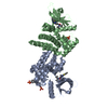
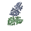
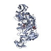
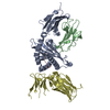
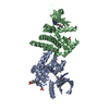
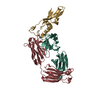
ACETATE ION / NICKEL (II) ION / PHOSPHATE ION / Bifunctional arginine demethylase and lysyl-hydroxylase JMJD6 Similarity search - Component
Biological species
Homo sapiens (human)
Method
X-RAY DIFFRACTION / SYNCHROTRON / SAD / Resolution: 1.75 Å
Mass: 39897.441 Da / Num. of mol.: 2 / Fragment: UNP RESIDUES 2-335 Source method: isolated from a genetically manipulated source Source: (gene. exp.) Homo sapiens (human) / Gene: JMJD6, KIAA0585, PTDSR / Production host: Escherichia coli (E. coli) References: UniProt: Q6NYC1, Oxidoreductases; Acting on paired donors, with incorporation or reduction of molecular oxygen; With 2-oxoglutarate as one donor, and incorporation of one atom of oxygen into each donor
In the structure databanks used in Yorodumi, some data are registered as the other names, "COVID-19 virus" and "2019-nCoV". Here are the details of the virus and the list of structure data.
Jan 31, 2019. EMDB accession codes are about to change! (news from PDBe EMDB page)
EMDB accession codes are about to change! (news from PDBe EMDB page)
The allocation of 4 digits for EMDB accession codes will soon come to an end. Whilst these codes will remain in use, new EMDB accession codes will include an additional digit and will expand incrementally as the available range of codes is exhausted. The current 4-digit format prefixed with “EMD-” (i.e. EMD-XXXX) will advance to a 5-digit format (i.e. EMD-XXXXX), and so on. It is currently estimated that the 4-digit codes will be depleted around Spring 2019, at which point the 5-digit format will come into force.
The EM Navigator/Yorodumi systems omit the EMD- prefix.
Related info.:Q: What is EMD? / ID/Accession-code notation in Yorodumi/EM Navigator
Yorodumi is a browser for structure data from EMDB, PDB, SASBDB, etc.
This page is also the successor to EM Navigator detail page, and also detail information page/front-end page for Omokage search.
The word "yorodu" (or yorozu) is an old Japanese word meaning "ten thousand". "mi" (miru) is to see.
Related info.:EMDB / PDB / SASBDB / Comparison of 3 databanks / Yorodumi Search / Aug 31, 2016. New EM Navigator & Yorodumi / Yorodumi Papers / Jmol/JSmol / Function and homology information / Changes in new EM Navigator and Yorodumi
 Movie
Movie Controller


 Open data
 Basic information
 Components
 Keywords
 Function and homology information
 Homo sapiens (human)
 X-RAY DIFFRACTION /
  Authors
 Citation
 Structure visualization
 Downloads & links
 Other downloads
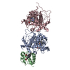


 PDBj
PDBj
 Assembly
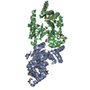


 Mass: 58.693 Da / Num. of mol.: 2 / Source method: obtained synthetically / Formula: Ni
 Mass: 22.990 Da / Num. of mol.: 2 / Source method: obtained synthetically / Formula: Na
 Mass: 35.453 Da / Num. of mol.: 6 / Source method: obtained synthetically / Formula: Cl
 Mass: 94.971 Da / Num. of mol.: 3 / Source method: obtained synthetically / Formula: PO4
 Mass: 59.044 Da / Num. of mol.: 2 / Source method: obtained synthetically / Formula: C2H3O2
 Mass: 92.094 Da / Num. of mol.: 1 / Source method: obtained synthetically / Formula: C3H8O3
 Sample preparation
 / Beamline: I04 / Wavelength: 0.9793 Å
 Processing