 Movie
Movie Controller

[English] 日本語
 Yorodumi
- PDB-3jxz: Bacillus cereus Alkylpurine DNA Glycosylase AlkD Bound to DNA Con... -
+ Open data

- Basic information
| Entry | Database: PDB / ID: 3jxz | ||||||
|---|---|---|---|---|---|---|---|
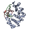
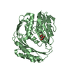
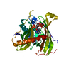
| Title | Bacillus cereus Alkylpurine DNA Glycosylase AlkD Bound to DNA Containing an Abasic Site (across from T) | ||||||
 Components Components |
| ||||||
 Keywords Keywords | hydrolase/DNA / HEAT repeat / DNA binding / DNA glycosylase / DNA alkylation / lyase-DNA COMPLEX / hydrolase-DNA COMPLEX | ||||||
| Function / homology |  Function and homology information Function and homology information | ||||||
| Biological species |  | ||||||
| Method |  X-RAY DIFFRACTION / SYNCHROTRON / MOLECULAR REPLACEMENT / Resolution: 1.75 Å X-RAY DIFFRACTION / SYNCHROTRON / MOLECULAR REPLACEMENT / Resolution: 1.75 Å | ||||||
 Authors Authors | Rubinson, E.H. / Eichman, B.F. | ||||||
 Citation Citation | Journal: Nature / Year: 2010 Title: An unprecedented nucleic acid capture mechanism for excision of DNA damage. Authors: Rubinson, E.H. / Gowda, A.S. / Spratt, T.E. / Gold, B. / Eichman, B.F. | ||||||
| History |
|
- Structure visualization
| Structure viewer | Molecule: MolmilJmol/JSmol |
|---|
- Downloads & links
-Download
| PDBx/mmCIF format | 3jxz.cif.gz | 135.7 KB | Display | PDBx/mmCIF format |
|---|---|---|---|---|
| PDB format | pdb3jxz.ent.gz | 101.8 KB | Display | PDB format |
| PDBx/mmJSON format | 3jxz.json.gz | Tree view | PDBx/mmJSON format | |
| Others |  Other downloads Other downloads |
-Validation report
| Arichive directory | https://data.pdbj.org/pub/pdb/validation_reports/jx/3jxzftp://data.pdbj.org/pub/pdb/validation_reports/jx/3jxz | HTTPS FTP |
|---|
-Related structure data
| Related structure data | 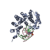 3jx7C 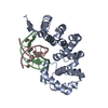 3jxyC  3jy1C  3bvsS S: Starting model for refinement C: citing same article ( |
|---|---|
| Similar structure data |


-Links
 PDBj
PDBj- Assembly
| Deposited unit | 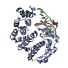
| ||||||||
|---|---|---|---|---|---|---|---|---|---|
| 1 |
| ||||||||
| Unit cell |
| ||||||||
| Details | biological unit = asymmetric unit |
-Components
| #1: Protein | Mass: 26970.260 Da / Num. of mol.: 1 Source method: isolated from a genetically manipulated source Source: (gene. exp.) |
|---|---|
| #2: DNA chain | Mass: 2982.927 Da / Num. of mol.: 1 / Source method: obtained synthetically |
| #3: DNA chain | Mass: 2973.971 Da / Num. of mol.: 1 / Source method: obtained synthetically |
| #4: Water | ChemComp-HOH /  Mass: 18.015 Da / Num. of mol.: 275 / Source method: isolated from a natural source / Formula: H2O Mass: 18.015 Da / Num. of mol.: 275 / Source method: isolated from a natural source / Formula: H2O |
-Experimental details
-Experiment
| Experiment | Method: X-RAY DIFFRACTION / Number of used crystals: 1 |
|---|
- Sample preparation
| Crystal | Density Matthews: 2.4 Å3/Da / Density % sol: 48.68 % |
|---|---|
| Crystal grow | Temperature: 289 K / Method: vapor diffusion, sitting drop / pH: 6.5 Details: .1 M BisTris pH 6.5 .1 M NaCl, 19% PEG 3350, 2% glycerol, VAPOR DIFFUSION, SITTING DROP, temperature 289K |
-Data collection
| Diffraction | Mean temperature: 100 K |
|---|---|
| Diffraction source | Source: SYNCHROTRON / Site: APS  / Beamline: 21-ID-D / Wavelength: 0.9785 Å / Beamline: 21-ID-D / Wavelength: 0.9785 Å |
| Detector | Type: MAR scanner 300 mm plate / Detector: IMAGE PLATE / Date: Aug 12, 2007 |
| Radiation | Monochromator: Si(111) / Protocol: SINGLE WAVELENGTH / Monochromatic (M) / Laue (L): M / Scattering type: x-ray |
| Radiation wavelength | Wavelength: 0.9785 Å / Relative weight: 1 |
| Reflection | Resolution: 1.75→28.9 Å / Num. all: 31562 / Num. obs: 30136 / % possible obs: 95.5 % / Observed criterion σ(F): 0 / Observed criterion σ(I): 0 / Redundancy: 3.3 % / Biso Wilson estimate: 22.4 Å2 / Rsym value: 0.056 / Net I/σ(I): 13.5 |
| Reflection shell | Resolution: 1.75→1.81 Å / Redundancy: 3.3 % / Mean I/σ(I) obs: 2.9 / Num. unique all: 2868 / Rsym value: 0.467 / % possible all: 94.8 |
- Processing
| Software |
| ||||||||||||||||||||||||||||||||||||||||||||||||||||||||||||||||||||||||||||||||||||||||||||||||||||||||
|---|---|---|---|---|---|---|---|---|---|---|---|---|---|---|---|---|---|---|---|---|---|---|---|---|---|---|---|---|---|---|---|---|---|---|---|---|---|---|---|---|---|---|---|---|---|---|---|---|---|---|---|---|---|---|---|---|---|---|---|---|---|---|---|---|---|---|---|---|---|---|---|---|---|---|---|---|---|---|---|---|---|---|---|---|---|---|---|---|---|---|---|---|---|---|---|---|---|---|---|---|---|---|---|---|---|
| Refinement | Method to determine structure: MOLECULAR REPLACEMENT Starting model: PDB 3BVS Resolution: 1.75→28.867 Å / SU ML: 0.12 / Cross valid method: THROUGHOUT / σ(F): 1.36 / Stereochemistry target values: ML
| ||||||||||||||||||||||||||||||||||||||||||||||||||||||||||||||||||||||||||||||||||||||||||||||||||||||||
| Solvent computation | Shrinkage radii: 0.9 Å / VDW probe radii: 1.11 Å / Solvent model: FLAT BULK SOLVENT MODEL / Bsol: 61.383 Å2 / ksol: 0.388 e/Å3 | ||||||||||||||||||||||||||||||||||||||||||||||||||||||||||||||||||||||||||||||||||||||||||||||||||||||||
| Displacement parameters | Biso mean: 32.42 Å2 | ||||||||||||||||||||||||||||||||||||||||||||||||||||||||||||||||||||||||||||||||||||||||||||||||||||||||
| Refinement step | Cycle: LAST / Resolution: 1.75→28.867 Å
| ||||||||||||||||||||||||||||||||||||||||||||||||||||||||||||||||||||||||||||||||||||||||||||||||||||||||
| Refine LS restraints |
| ||||||||||||||||||||||||||||||||||||||||||||||||||||||||||||||||||||||||||||||||||||||||||||||||||||||||
| LS refinement shell |
| ||||||||||||||||||||||||||||||||||||||||||||||||||||||||||||||||||||||||||||||||||||||||||||||||||||||||
| Refinement TLS params. | Refine-ID: X-RAY DIFFRACTION
| ||||||||||||||||||||||||||||||||||||||||||||||||||||||||||||||||||||||||||||||||||||||||||||||||||||||||
| Refinement TLS group | Selection details: CHAIN C |
