 Movie
Movie Controller


+ Open data

- Basic information
| Entry | Database: PDB / ID: 3jtt | ||||||
|---|---|---|---|---|---|---|---|
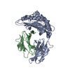
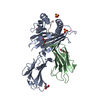
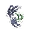
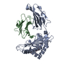
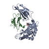
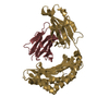
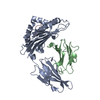
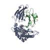
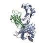
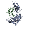
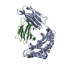
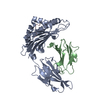
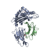
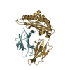
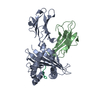
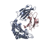
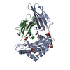
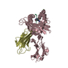
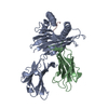
| Title | Cystal structure of Rhesus macaque MHC class I:Mamu-A*02 | ||||||
 Components Components |
| ||||||
 Keywords Keywords | IMMUNE SYSTEM / ALPHA HELIX / BETA SHEET / BETA BARREL / Immune response / MHC I / Membrane / Transmembrane / Disease mutation / Disulfide bond / Glycation / Glycoprotein / Immunoglobulin domain / Pyrrolidone carboxylic acid / Secreted | ||||||
| Function / homology |  Function and homology information Function and homology informationantigen processing and presentation of peptide antigen via MHC class I / T cell mediated cytotoxicity / lumenal side of endoplasmic reticulum membrane / cellular response to iron(III) ion / negative regulation of iron ion transport / negative regulation of forebrain neuron differentiation / antigen processing and presentation of exogenous protein antigen via MHC class Ib, TAP-dependent / peptide antigen assembly with MHC class I protein complex / regulation of erythrocyte differentiation / ER to Golgi transport vesicle membrane ...antigen processing and presentation of peptide antigen via MHC class I / T cell mediated cytotoxicity / lumenal side of endoplasmic reticulum membrane / cellular response to iron(III) ion / negative regulation of iron ion transport / negative regulation of forebrain neuron differentiation / antigen processing and presentation of exogenous protein antigen via MHC class Ib, TAP-dependent / peptide antigen assembly with MHC class I protein complex / regulation of erythrocyte differentiation / ER to Golgi transport vesicle membrane / response to molecule of bacterial origin / HFE-transferrin receptor complex / MHC class I peptide loading complex / transferrin transport / negative regulation of receptor-mediated endocytosis / positive regulation of T cell cytokine production / antigen processing and presentation of endogenous peptide antigen via MHC class I / MHC class I protein complex / peptide antigen assembly with MHC class II protein complex / negative regulation of neurogenesis / cellular response to nicotine / MHC class II protein complex / positive regulation of receptor-mediated endocytosis / multicellular organismal-level iron ion homeostasis / positive regulation of T cell mediated cytotoxicity / antigen processing and presentation of exogenous peptide antigen via MHC class II / positive regulation of immune response / peptide antigen binding / phagocytic vesicle membrane / recycling endosome membrane / negative regulation of epithelial cell proliferation / positive regulation of T cell activation / sensory perception of smell / positive regulation of cellular senescence / MHC class II protein complex binding / T cell differentiation in thymus / late endosome membrane / negative regulation of neuron projection development / protein refolding / early endosome membrane / amyloid fibril formation / protein homotetramerization / intracellular iron ion homeostasis / learning or memory / immune response / Golgi membrane / external side of plasma membrane / lysosomal membrane / GTP binding / structural molecule activity / cell surface / Golgi apparatus / protein homodimerization activity / extracellular region / cytosol Similarity search - Function | ||||||
| Biological species |  | ||||||
| Method |  X-RAY DIFFRACTION / MOLECULAR REPLACEMENT / Resolution: 2.8 Å X-RAY DIFFRACTION / MOLECULAR REPLACEMENT / Resolution: 2.8 Å | ||||||
 Authors Authors | Dai, L. / Feng, Y. / Qi, J. / Gao, G.F. | ||||||
 Citation Citation | Journal: To be Published Title: Crystal structure of Rhesus macaque MHC class I:Mamu-A*02 Authors: Dai, L. | ||||||
| History |
|
- Structure visualization
| Structure viewer | Molecule: MolmilJmol/JSmol |
|---|
- Downloads & links
-Download
| PDBx/mmCIF format | 3jtt.cif.gz | 247.5 KB | Display | PDBx/mmCIF format |
|---|---|---|---|---|
| PDB format | pdb3jtt.ent.gz | 201.7 KB | Display | PDB format |
| PDBx/mmJSON format | 3jtt.json.gz | Tree view | PDBx/mmJSON format | |
| Others |  Other downloads Other downloads |
-Validation report
| Arichive directory | https://data.pdbj.org/pub/pdb/validation_reports/jt/3jttftp://data.pdbj.org/pub/pdb/validation_reports/jt/3jtt | HTTPS FTP |
|---|
-Related structure data
| Related structure data |  1zvsS S: Starting model for refinement |
|---|---|
| Similar structure data |


-Links
 PDBj
PDBj

- Assembly
| Deposited unit | 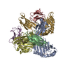
| ||||||||
|---|---|---|---|---|---|---|---|---|---|
| 1 | 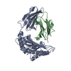
| ||||||||
| 2 | 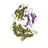
| ||||||||
| 3 | 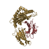
| ||||||||
| Unit cell |
|
-Components
| #1: Protein | Mass: 32106.320 Da / Num. of mol.: 3 / Fragment: UNP residues 17-292 Source method: isolated from a genetically manipulated source Source: (gene. exp.)  #2: Protein | Mass: 11791.275 Da / Num. of mol.: 3 Source method: isolated from a genetically manipulated source Source: (gene. exp.) #3: Protein/peptide | Mass: 1014.114 Da / Num. of mol.: 3 / Fragment: UNP residues 159-167 / Source method: obtained synthetically Details: This sequence occurs from Simian immunodeficiency virus References: UniProt: Q9WH73 #4: Water | ChemComp-HOH / |  Mass: 18.015 Da / Num. of mol.: 270 / Source method: isolated from a natural source / Formula: H2O Mass: 18.015 Da / Num. of mol.: 270 / Source method: isolated from a natural source / Formula: H2OHas protein modification | Y | |
|---|
-Experimental details
-Experiment
| Experiment | Method: X-RAY DIFFRACTION / Number of used crystals: 1 |
|---|
- Sample preparation
| Crystal | Density Matthews: 3.98 Å3/Da / Density % sol: 69.13 % |
|---|---|
| Crystal grow | Temperature: 291 K / Method: vapor diffusion, hanging drop / pH: 5.5 Details: 0.1 M Bis-Tris pH 5.5, 2 M ammonium sulfate , VAPOR DIFFUSION, HANGING DROP, temperature 291K |
-Data collection
| Diffraction | Mean temperature: 100 K |
|---|---|
| Diffraction source | Source: ROTATING ANODE / Type: RIGAKU MICROMAX-007 HF / Wavelength: 1.5478 Å |
| Detector | Type: RIGAKU RAXIS IV++ / Detector: IMAGE PLATE / Date: May 20, 2008 / Details: mirrors |
| Radiation | Monochromator: GRAPHITE / Protocol: SINGLE WAVELENGTH / Monochromatic (M) / Laue (L): M / Scattering type: x-ray |
| Radiation wavelength | Wavelength: 1.5478 Å / Relative weight: 1 |
| Reflection | Resolution: 2.8→50 Å / Num. obs: 51015 / % possible obs: 99.6 % / Observed criterion σ(F): 2 / Observed criterion σ(I): 2 / Redundancy: 7.1 % / Biso Wilson estimate: 57 Å2 / Rmerge(I) obs: 0.074 / Rsym value: 0.074 / Net I/σ(I): 37.2 |
| Reflection shell | Resolution: 2.8→2.9 Å / Redundancy: 7 % / Rmerge(I) obs: 0.528 / Mean I/σ(I) obs: 4.7 / Num. unique all: 4987 / Rsym value: 0.528 / % possible all: 100 |
- Processing
| Software |
| |||||||||||||||||||||||||||||||||||||||||||||||||||||||||||||||||||||||||||||||||||||||||||||||||||||||||||||||||||||||||||||||||||||
|---|---|---|---|---|---|---|---|---|---|---|---|---|---|---|---|---|---|---|---|---|---|---|---|---|---|---|---|---|---|---|---|---|---|---|---|---|---|---|---|---|---|---|---|---|---|---|---|---|---|---|---|---|---|---|---|---|---|---|---|---|---|---|---|---|---|---|---|---|---|---|---|---|---|---|---|---|---|---|---|---|---|---|---|---|---|---|---|---|---|---|---|---|---|---|---|---|---|---|---|---|---|---|---|---|---|---|---|---|---|---|---|---|---|---|---|---|---|---|---|---|---|---|---|---|---|---|---|---|---|---|---|---|---|---|
| Refinement | Method to determine structure: MOLECULAR REPLACEMENT Starting model: PDB ENTRY 1zvs Resolution: 2.8→31.293 Å / SU ML: 0.4 / Isotropic thermal model: Isotropic / Cross valid method: THROUGHOUT / σ(F): 0.09 / Stereochemistry target values: ML / Details: Used weighted full matrix least squares procedure
| |||||||||||||||||||||||||||||||||||||||||||||||||||||||||||||||||||||||||||||||||||||||||||||||||||||||||||||||||||||||||||||||||||||
| Solvent computation | Shrinkage radii: 0.9 Å / VDW probe radii: 1.11 Å / Solvent model: FLAT BULK SOLVENT MODEL / Bsol: 45.463 Å2 / ksol: 0.356 e/Å3 | |||||||||||||||||||||||||||||||||||||||||||||||||||||||||||||||||||||||||||||||||||||||||||||||||||||||||||||||||||||||||||||||||||||
| Displacement parameters | Biso mean: 57.9 Å2
| |||||||||||||||||||||||||||||||||||||||||||||||||||||||||||||||||||||||||||||||||||||||||||||||||||||||||||||||||||||||||||||||||||||
| Refinement step | Cycle: LAST / Resolution: 2.8→31.293 Å
| |||||||||||||||||||||||||||||||||||||||||||||||||||||||||||||||||||||||||||||||||||||||||||||||||||||||||||||||||||||||||||||||||||||
| Refine LS restraints |
| |||||||||||||||||||||||||||||||||||||||||||||||||||||||||||||||||||||||||||||||||||||||||||||||||||||||||||||||||||||||||||||||||||||
| LS refinement shell |
|
