[English] 日本語
 Yorodumi
- PDB-3jaa: HUMAN DNA POLYMERASE ETA in COMPLEX WITH NORMAL DNA AND INCO NUCL... -
+ Open data
- Basic information
| Entry | Database: PDB / ID: 3jaa | ||||||
|---|---|---|---|---|---|---|---|
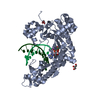
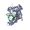
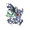
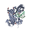
| Title | HUMAN DNA POLYMERASE ETA in COMPLEX WITH NORMAL DNA AND INCO NUCLEOTIDE (NRM) | ||||||
 Components Components |
| ||||||
 Keywords Keywords | TRANSFERASE/DNA / POL ETA / POLYMERASE / THYMINE DIMER / CPD / XPV / XERODERMA PIGM VARIANT / DNA DAMAGE / TRANSFERASE-DNA COMPLEX | ||||||
| Function / homology |  Function and homology information Function and homology informationresponse to UV-C / error-free translesion synthesis / DNA synthesis involved in DNA repair / cellular response to UV-C / pyrimidine dimer repair / error-prone translesion synthesis / regulation of DNA repair / Termination of translesion DNA synthesis / replication fork / Translesion Synthesis by POLH ...response to UV-C / error-free translesion synthesis / DNA synthesis involved in DNA repair / cellular response to UV-C / pyrimidine dimer repair / error-prone translesion synthesis / regulation of DNA repair / Termination of translesion DNA synthesis / replication fork / Translesion Synthesis by POLH / HDR through Homologous Recombination (HRR) / response to radiation / site of double-strand break / damaged DNA binding / DNA-directed DNA polymerase / DNA-directed DNA polymerase activity / DNA replication / DNA repair / zinc ion binding / nucleoplasm / nucleus / cytosol Similarity search - Function | ||||||
| Biological species |  Homo sapiens (human) Homo sapiens (human)synthetic construct (others) | ||||||
| Method | ELECTRON MICROSCOPY / single particle reconstruction / negative staining / Resolution: 22 Å | ||||||
 Authors Authors | Lau, W.C.Y. / Li, Y. / Zhang, Q. / Huen, M.S.Y. | ||||||
 Citation Citation |  Journal: Sci Rep / Year: 2015 Journal: Sci Rep / Year: 2015Title: Molecular architecture of the Ub-PCNA/Pol η complex bound to DNA. Authors: Wilson C Y Lau / Yinyin Li / Qinfen Zhang / Michael S Y Huen /  Abstract: Translesion synthesis (TLS) is the mechanism by which DNA polymerases replicate through unrepaired DNA lesions. TLS is activated by monoubiquitination of the homotrimeric proliferating cell nuclear ...Translesion synthesis (TLS) is the mechanism by which DNA polymerases replicate through unrepaired DNA lesions. TLS is activated by monoubiquitination of the homotrimeric proliferating cell nuclear antigen (PCNA) at lysine-164, followed by the switch from replicative to specialized polymerases at DNA damage sites. Pol η belongs to the Y-Family of specialized polymerases that can efficiently bypass UV-induced lesions. Like other members of the Y-Family polymerases, its recruitment to the damaged sites is mediated by the interaction with monoubiquitinated PCNA (Ub-PCNA) via its ubiquitin-binding domain and non-canonical PCNA-interacting motif in the C-terminal region. The structural determinants underlying the direct recognition of Ub-PCNA by Pol η, or Y-Family polymerases in general, remain largely unknown. Here we report a structure of the Ub-PCNA/Pol η complex bound to DNA determined by single-particle electron microscopy (EM). The overall obtained structure resembles that of the editing PCNA/PolB complex. Analysis of the map revealed the conformation of ubiquitin that binds the C-terminal domain of Pol η. Our present study suggests that the Ub-PCNA/Pol η interaction requires the formation of a structured binding interface, which is dictated by the inherent flexibility of Ub-PCNA. | ||||||
| History |
|
- Structure visualization
| Movie |
 Movie viewer Movie viewer |
|---|---|
| Structure viewer | Molecule:  Molmil Molmil Jmol/JSmol Jmol/JSmol |
- Downloads & links
- Download
| PDBx/mmCIF format |  3jaa.cif.gz 3jaa.cif.gz | 129.1 KB | Display |  PDBx/mmCIF format PDBx/mmCIF format |
|---|---|---|---|---|
| PDB format |  pdb3jaa.ent.gz pdb3jaa.ent.gz | 93.5 KB | Display |  PDB format PDB format |
| PDBx/mmJSON format |  3jaa.json.gz 3jaa.json.gz | Tree view |  PDBx/mmJSON format PDBx/mmJSON format | |
| Others |  Other downloads Other downloads |
-Validation report
| Summary document |  3jaa_validation.pdf.gz 3jaa_validation.pdf.gz | 897.9 KB | Display |  wwPDB validaton report wwPDB validaton report |
|---|---|---|---|---|
| Full document |  3jaa_full_validation.pdf.gz 3jaa_full_validation.pdf.gz | 899 KB | Display | |
| Data in XML |  3jaa_validation.xml.gz 3jaa_validation.xml.gz | 21.9 KB | Display | |
| Data in CIF |  3jaa_validation.cif.gz 3jaa_validation.cif.gz | 34.1 KB | Display | |
| Arichive directory |  https://data.pdbj.org/pub/pdb/validation_reports/ja/3jaa https://data.pdbj.org/pub/pdb/validation_reports/ja/3jaa ftp://data.pdbj.org/pub/pdb/validation_reports/ja/3jaa ftp://data.pdbj.org/pub/pdb/validation_reports/ja/3jaa | HTTPS FTP |
-Related structure data
| Related structure data |  6339MC  3ja9C M: map data used to model this data C: citing same article ( |
|---|---|
| Similar structure data |
- Links
Links
- Assembly
| Deposited unit | 
|
|---|---|
| 1 |
|
- Components
-Protein , 1 types, 1 molecules A
| #1: Protein | Mass: 48617.707 Da / Num. of mol.: 1 / Fragment: CATALYTIC CORE (RESIDUES 1-432) Source method: isolated from a genetically manipulated source Source: (gene. exp.)  Homo sapiens (human) / Gene: POLH, RAD30, RAD30A, XPV / Plasmid: PET28 / Production host: Homo sapiens (human) / Gene: POLH, RAD30, RAD30A, XPV / Plasmid: PET28 / Production host:  |
|---|
-DNA chain , 2 types, 2 molecules TP
| #2: DNA chain | Mass: 3941.584 Da / Num. of mol.: 1 / Fragment: DNA TEMPLATE / Source method: obtained synthetically / Source: (synth.) synthetic construct (others) |
|---|---|
| #3: DNA chain | Mass: 2730.810 Da / Num. of mol.: 1 / Fragment: DNA PRIMER / Source method: obtained synthetically / Source: (synth.) synthetic construct (others) |
-Non-polymers , 4 types, 412 molecules 


| #4: Chemical | | #5: Chemical | #6: Chemical | ChemComp-GOL / #7: Water | ChemComp-HOH / | |
|---|
-Experimental details
-Experiment
| Experiment | Method: ELECTRON MICROSCOPY / Number of used crystals: 1 |
|---|---|
| EM experiment | Aggregation state: PARTICLE / 3D reconstruction method: single particle reconstruction |
- Sample preparation
| Component | Name: Ub-PCNA/Pol eta/DNA / Type: COMPLEX Details: Monomeric catalytic core of Pol eta binds to one homotrimeric Ub-PCNA |
|---|---|
| Molecular weight | Value: 0.2 MDa / Experimental value: YES |
| Buffer solution | pH: 8 / Details: 50 mM Tris-HCl, 5 mM MgCl2 |
| Specimen | Embedding applied: NO / Shadowing applied: NO / Staining applied: YES / Vitrification applied: NO Details: 50 mM Tris-HCl, 5 mM MgCl2 (Stain Details Grids with adsorbed protein floated on two 20 ul drops of 2% w/v uranyl acetate solution for 30 seconds) |
| EM staining | Type: NEGATIVE / Material: Uranyl Acetate |
| Specimen support | Details: Continuous carbon coated copper grids (Ted Pella), glow-discharged for 30 seconds |
- Electron microscopy imaging
| Microscopy | Model: JEOL 2010 / Date: Nov 12, 2014 |
|---|---|
| Electron gun | Electron source: LAB6 / Accelerating voltage: 200 kV / Illumination mode: FLOOD BEAM |
| Electron lens | Mode: BRIGHT FIELD / Nominal magnification: 50000 X / Camera length: 0 mm |
| Specimen holder | Specimen holder model: SIDE ENTRY, EUCENTRIC / Tilt angle max: 0 ° / Tilt angle min: 0 ° |
| Image recording | Electron dose: 18 e/Å2 / Film or detector model: GATAN ULTRASCAN 4000 (4k x 4k) |
- Processing
| EM software |
| ||||||||||||
|---|---|---|---|---|---|---|---|---|---|---|---|---|---|
| CTF correction | Details: Particles | ||||||||||||
| Symmetry | Point symmetry: C1 (asymmetric) | ||||||||||||
| 3D reconstruction | Method: Projection matching / Resolution: 22 Å / Resolution method: FSC 0.143 CUT-OFF / Num. of particles: 7330 / Details: (Single particle--Applied symmetry: C1) / Num. of class averages: 227 / Symmetry type: POINT | ||||||||||||
| Atomic model building | Protocol: RIGID BODY FIT / Space: REAL / Details: REFINEMENT PROTOCOL--RIGID BODY | ||||||||||||
| Atomic model building | PDB-ID: 3MR2 Accession code: 3MR2 / Source name: PDB / Type: experimental model |
 Movie
Movie Controller


 UCSF Chimera


 PDBj
PDBj


