 Movie
Movie Controller

[English] 日本語
 Yorodumi
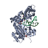
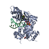
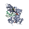
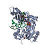
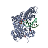
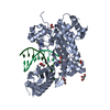
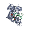
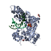
- PDB-5dqg: Crystal Structure of Human DNA Polymerase Eta Inserting dAMPNPP O... -
+ Open data

- Basic information
| Entry | Database: PDB / ID: 5dqg | ||||||
|---|---|---|---|---|---|---|---|
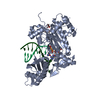
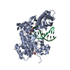
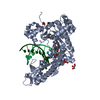
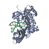
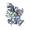
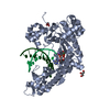
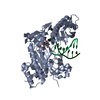
| Title | Crystal Structure of Human DNA Polymerase Eta Inserting dAMPNPP Opposite O4-Ethylthymidine | ||||||
 Components Components |
| ||||||
 Keywords Keywords | Transferase/DNA / Catalytic Domain / DNA Damage / DNA-Directed DNA Polymerase / Adenosine Triphosphate / Y-family polymerase / trans-lesion synthesis (TLS) / DNA Binding / O4-Alkylthymidine / O4-Ethylthymidine / Transferase-DNA complex | ||||||
| Function / homology |  Function and homology information Function and homology informationresponse to UV-C / error-free translesion synthesis / DNA synthesis involved in DNA repair / cellular response to UV-C / pyrimidine dimer repair / error-prone translesion synthesis / regulation of DNA repair / replication fork / Termination of translesion DNA synthesis / response to radiation ...response to UV-C / error-free translesion synthesis / DNA synthesis involved in DNA repair / cellular response to UV-C / pyrimidine dimer repair / error-prone translesion synthesis / regulation of DNA repair / replication fork / Termination of translesion DNA synthesis / response to radiation / Translesion Synthesis by POLH / HDR through Homologous Recombination (HRR) / site of double-strand break / DNA-directed DNA polymerase / damaged DNA binding / DNA-directed DNA polymerase activity / DNA replication / DNA repair / zinc ion binding / nucleoplasm / nucleus / cytosol Similarity search - Function | ||||||
| Biological species |  Homo sapiens (human) Homo sapiens (human)synthetic construct (others) | ||||||
| Method |  X-RAY DIFFRACTION / SYNCHROTRON / MOLECULAR REPLACEMENT / Resolution: 2.29 Å X-RAY DIFFRACTION / SYNCHROTRON / MOLECULAR REPLACEMENT / Resolution: 2.29 Å | ||||||
 Authors Authors | Patra, A. / OFlaherty, D.K. / Egli, M. | ||||||
 Citation Citation | Journal: Chem Sci / Year: 2016 Title: Lesion Orientation ofO4-Alkylthymidine Influences Replication by Human DNA Polymeraseeta. Authors: O'Flaherty, D.K. / Patra, A. / Su, Y. / Guengerich, F.P. / Egli, M. / Wilds, C.J. | ||||||
| History |
|
- Structure visualization
| Structure viewer | Molecule: MolmilJmol/JSmol |
|---|
- Downloads & links
-Download
| PDBx/mmCIF format | 5dqg.cif.gz | 121.2 KB | Display | PDBx/mmCIF format |
|---|---|---|---|---|
| PDB format | pdb5dqg.ent.gz | 86.3 KB | Display | PDB format |
| PDBx/mmJSON format | 5dqg.json.gz | Tree view | PDBx/mmJSON format | |
| Others |  Other downloads Other downloads |
-Validation report
| Arichive directory | https://data.pdbj.org/pub/pdb/validation_reports/dq/5dqgftp://data.pdbj.org/pub/pdb/validation_reports/dq/5dqg | HTTPS FTP |
|---|
-Related structure data
| Related structure data |  5dlfC  5dlgC  5dqhC  5dqiC  4o3nS C: citing same article ( S: Starting model for refinement |
|---|---|
| Similar structure data |


-Links
 PDBj
PDBj


- Assembly
| Deposited unit | 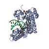
| ||||||||
|---|---|---|---|---|---|---|---|---|---|
| 1 |
| ||||||||
| Unit cell |
|
-Components
-Protein , 1 types, 1 molecules A
| #1: Protein | Mass: 48617.707 Da / Num. of mol.: 1 / Fragment: UNP residues 1-432 Source method: isolated from a genetically manipulated source Source: (gene. exp.) Homo sapiens (human) / Gene: POLH, RAD30, RAD30A, XPV / Production host:  |
|---|
-DNA chain , 2 types, 2 molecules TP
| #2: DNA chain | Mass: 3665.445 Da / Num. of mol.: 1 / Source method: obtained synthetically / Details: Chemically Synthesized / Source: (synth.) synthetic construct (others) |
|---|---|
| #3: DNA chain | Mass: 2426.617 Da / Num. of mol.: 1 / Source method: obtained synthetically / Details: Chemically Synthesized / Source: (synth.) synthetic construct (others) |
-Non-polymers , 4 types, 216 molecules 


| #4: Chemical | ChemComp-DZ4 /  Mass: 490.197 Da / Num. of mol.: 1 / Source method: obtained synthetically / Formula: C10H17N6O11P3 Mass: 490.197 Da / Num. of mol.: 1 / Source method: obtained synthetically / Formula: C10H17N6O11P3 | ||||
|---|---|---|---|---|---|
| #5: Chemical |  Mass: 24.305 Da / Num. of mol.: 2 / Source method: obtained synthetically / Formula: Mg Mass: 24.305 Da / Num. of mol.: 2 / Source method: obtained synthetically / Formula: Mg#6: Chemical | ChemComp-GOL / |  Mass: 92.094 Da / Num. of mol.: 1 / Source method: obtained synthetically / Formula: C3H8O3 Mass: 92.094 Da / Num. of mol.: 1 / Source method: obtained synthetically / Formula: C3H8O3#7: Water | ChemComp-HOH / | Mass: 18.015 Da / Num. of mol.: 212 / Source method: isolated from a natural source / Formula: H2O |
-Experimental details
-Experiment
| Experiment | Method: X-RAY DIFFRACTION |
|---|
- Sample preparation
| Crystal | Density Matthews: 2.11 Å3/Da / Density % sol: 41.79 % |
|---|---|
| Crystal grow | Temperature: 291 K / Method: vapor diffusion, hanging drop / pH: 5.5 Details: 0.1 M MES pH 5.5, 5mM magnesium chloride, 22% PEG 2000 MME |
-Data collection
| Diffraction | Mean temperature: 100 K |
|---|---|
| Diffraction source | Source: SYNCHROTRON / Site: APS  / Beamline: 21-ID-G / Wavelength: 0.97856 Å / Beamline: 21-ID-G / Wavelength: 0.97856 Å |
| Detector | Type: MARMOSAIC 300 mm CCD / Detector: CCD / Date: Aug 15, 2014 |
| Radiation | Monochromator: C(111) / Protocol: SINGLE WAVELENGTH / Monochromatic (M) / Laue (L): M / Scattering type: x-ray |
| Radiation wavelength | Wavelength: 0.97856 Å / Relative weight: 1 |
| Reflection | Resolution: 2.29→59.21 Å / Num. all: 20600 / Num. obs: 20600 / % possible obs: 100 % / Observed criterion σ(F): 0 / Observed criterion σ(I): -3 / Redundancy: 5.6 % / Rmerge(I) obs: 0.155 / Rsym value: 0.155 / Net I/σ(I): 8.4 |
| Reflection shell | Resolution: 2.29→2.35 Å / Redundancy: 5.6 % / Rmerge(I) obs: 0.595 / Mean I/σ(I) obs: 3.2 / % possible all: 100 |
- Processing
| Software |
| ||||||||||||||||||||||||||||||||||||||||||||||||||||||||||||||||||||||||||||||||||||||||||||||||||||||||||||||||||||||||||||||||||||||||||||||||||||||||||||||||||||||||||||||||||||||
|---|---|---|---|---|---|---|---|---|---|---|---|---|---|---|---|---|---|---|---|---|---|---|---|---|---|---|---|---|---|---|---|---|---|---|---|---|---|---|---|---|---|---|---|---|---|---|---|---|---|---|---|---|---|---|---|---|---|---|---|---|---|---|---|---|---|---|---|---|---|---|---|---|---|---|---|---|---|---|---|---|---|---|---|---|---|---|---|---|---|---|---|---|---|---|---|---|---|---|---|---|---|---|---|---|---|---|---|---|---|---|---|---|---|---|---|---|---|---|---|---|---|---|---|---|---|---|---|---|---|---|---|---|---|---|---|---|---|---|---|---|---|---|---|---|---|---|---|---|---|---|---|---|---|---|---|---|---|---|---|---|---|---|---|---|---|---|---|---|---|---|---|---|---|---|---|---|---|---|---|---|---|---|---|
| Refinement | Method to determine structure: MOLECULAR REPLACEMENT Starting model: 4O3N Resolution: 2.29→59.21 Å / Cor.coef. Fo:Fc: 0.943 / Cor.coef. Fo:Fc free: 0.919 / SU B: 7.528 / SU ML: 0.183 / Cross valid method: THROUGHOUT / ESU R: 0.441 / ESU R Free: 0.24 / Stereochemistry target values: MAXIMUM LIKELIHOOD / Details: HYDROGENS HAVE BEEN ADDED IN THE RIDING POSITIONS
| ||||||||||||||||||||||||||||||||||||||||||||||||||||||||||||||||||||||||||||||||||||||||||||||||||||||||||||||||||||||||||||||||||||||||||||||||||||||||||||||||||||||||||||||||||||||
| Solvent computation | Ion probe radii: 0.8 Å / Shrinkage radii: 0.8 Å / VDW probe radii: 1.2 Å / Solvent model: MASK | ||||||||||||||||||||||||||||||||||||||||||||||||||||||||||||||||||||||||||||||||||||||||||||||||||||||||||||||||||||||||||||||||||||||||||||||||||||||||||||||||||||||||||||||||||||||
| Displacement parameters | Biso mean: 31.071 Å2
| ||||||||||||||||||||||||||||||||||||||||||||||||||||||||||||||||||||||||||||||||||||||||||||||||||||||||||||||||||||||||||||||||||||||||||||||||||||||||||||||||||||||||||||||||||||||
| Refinement step | Cycle: 1 / Resolution: 2.29→59.21 Å
| ||||||||||||||||||||||||||||||||||||||||||||||||||||||||||||||||||||||||||||||||||||||||||||||||||||||||||||||||||||||||||||||||||||||||||||||||||||||||||||||||||||||||||||||||||||||
| Refine LS restraints |
|