Movie
Movie Controller

[English] 日本語
 Yorodumi
- PDB-3j8x: High-resolution structure of no-nucleotide kinesin on microtubules -
+ Open data

- Basic information
| Entry | Database: PDB / ID: 3j8x | ||||||
|---|---|---|---|---|---|---|---|
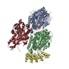
| Title | High-resolution structure of no-nucleotide kinesin on microtubules | ||||||
 Components Components |
| ||||||
 Keywords Keywords | MOTOR PROTEIN/STRUCTURAL PROTEIN / molecular motors / kinesin / myosin / microtubules / cytoskeletal motors / MOTOR PROTEIN-STRUCTURAL PROTEIN complex | ||||||
| Function / homology |  Function and homology information Function and homology informationregulation of modification of synapse structure, modulating synaptic transmission / plus-end-directed vesicle transport along microtubule / cytoplasm organization / cytolytic granule membrane / anterograde dendritic transport of neurotransmitter receptor complex / anterograde neuronal dense core vesicle transport / mitocytosis / retrograde neuronal dense core vesicle transport / anterograde axonal protein transport / ciliary rootlet ...regulation of modification of synapse structure, modulating synaptic transmission / plus-end-directed vesicle transport along microtubule / cytoplasm organization / cytolytic granule membrane / anterograde dendritic transport of neurotransmitter receptor complex / anterograde neuronal dense core vesicle transport / mitocytosis / retrograde neuronal dense core vesicle transport / anterograde axonal protein transport / ciliary rootlet / lysosome localization / positive regulation of potassium ion transport / plus-end-directed microtubule motor activity / vesicle transport along microtubule / RHO GTPases activate KTN1 / Kinesins / Microtubule-dependent trafficking of connexons from Golgi to the plasma membrane / Resolution of Sister Chromatid Cohesion / Hedgehog 'off' state / Cilium Assembly / Intraflagellar transport / COPI-dependent Golgi-to-ER retrograde traffic / Mitotic Prometaphase / Carboxyterminal post-translational modifications of tubulin / RHOH GTPase cycle / EML4 and NUDC in mitotic spindle formation / Sealing of the nuclear envelope (NE) by ESCRT-III / Kinesins / PKR-mediated signaling / Separation of Sister Chromatids / The role of GTSE1 in G2/M progression after G2 checkpoint / Aggrephagy / kinesin complex / RHO GTPases activate IQGAPs / RHO GTPases Activate Formins / HSP90 chaperone cycle for steroid hormone receptors (SHR) in the presence of ligand / microtubule motor activity / MHC class II antigen presentation / Recruitment of NuMA to mitotic centrosomes / centrosome localization / mitochondrion transport along microtubule / COPI-mediated anterograde transport / COPI-dependent Golgi-to-ER retrograde traffic / microtubule-based movement / stress granule disassembly / natural killer cell mediated cytotoxicity / Insulin processing / synaptic vesicle transport / postsynaptic cytosol / microtubule-based process / phagocytic vesicle / axon cytoplasm / MHC class II antigen presentation / dendrite cytoplasm / axon guidance / positive regulation of synaptic transmission, GABAergic / regulation of membrane potential / positive regulation of protein localization to plasma membrane / cellular response to type II interferon / structural constituent of cytoskeleton / microtubule cytoskeleton organization / centriolar satellite / Signaling by ALK fusions and activated point mutants / mitotic cell cycle / microtubule cytoskeleton / nuclear membrane / microtubule binding / vesicle / Hydrolases; Acting on acid anhydrides; Acting on GTP to facilitate cellular and subcellular movement / microtubule / cadherin binding / GTPase activity / GTP binding / protein-containing complex binding / perinuclear region of cytoplasm / ATP hydrolysis activity / mitochondrion / ATP binding / metal ion binding / identical protein binding / membrane / cytoplasm / cytosol Similarity search - Function | ||||||
| Biological species |  Homo sapiens (human) Homo sapiens (human) | ||||||
| Method | ELECTRON MICROSCOPY / helical reconstruction / cryo EM / Resolution: 5 Å | ||||||
 Authors Authors | Shang, Z. / Zhou, K. / Xu, C. / Csencsits, R. / Cochran, J.C. / Sindelar, C.V. | ||||||
 Citation Citation | Journal: Elife / Year: 2014 Title: High-resolution structures of kinesin on microtubules provide a basis for nucleotide-gated force-generation. Authors: Zhiguo Shang / Kaifeng Zhou / Chen Xu / Roseann Csencsits / Jared C Cochran / Charles V Sindelar /  Abstract: Microtubule-based transport by the kinesin motors, powered by ATP hydrolysis, is essential for a wide range of vital processes in eukaryotes. We obtained insight into this process by developing ...Microtubule-based transport by the kinesin motors, powered by ATP hydrolysis, is essential for a wide range of vital processes in eukaryotes. We obtained insight into this process by developing atomic models for no-nucleotide and ATP states of the monomeric kinesin motor domain on microtubules from cryo-EM reconstructions at 5-6 Å resolution. By comparing these models with existing X-ray structures of ADP-bound kinesin, we infer a mechanistic scheme in which microtubule attachment, mediated by a universally conserved 'linchpin' residue in kinesin (N255), triggers a clamshell opening of the nucleotide cleft and accompanying release of ADP. Binding of ATP re-closes the cleft in a manner that tightly couples to translocation of cargo, via kinesin's 'neck linker' element. These structural transitions are reminiscent of the analogous nucleotide-exchange steps in the myosin and F1-ATPase motors and inform how the two heads of a kinesin dimer 'gate' each other to promote coordinated stepping along microtubules. | ||||||
| History |
|
- Structure visualization
| Movie |
Movie viewer |
|---|---|
| Structure viewer | Molecule: MolmilJmol/JSmol |
- Downloads & links
-Download
| PDBx/mmCIF format | 3j8x.cif.gz | 221.7 KB | Display | PDBx/mmCIF format |
|---|---|---|---|---|
| PDB format | pdb3j8x.ent.gz | 166.1 KB | Display | PDB format |
| PDBx/mmJSON format | 3j8x.json.gz | Tree view | PDBx/mmJSON format | |
| Others |  Other downloads Other downloads |
-Validation report
| Summary document | 3j8x_validation.pdf.gz | 1 MB | Display | wwPDB validaton report |
|---|---|---|---|---|
| Full document | 3j8x_full_validation.pdf.gz | 1.1 MB | Display | |
| Data in XML | 3j8x_validation.xml.gz | 40.3 KB | Display | |
| Data in CIF | 3j8x_validation.cif.gz | 59.8 KB | Display | |
| Arichive directory | https://data.pdbj.org/pub/pdb/validation_reports/j8/3j8xftp://data.pdbj.org/pub/pdb/validation_reports/j8/3j8x | HTTPS FTP |
-Related structure data
| Related structure data | 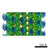 6187MC 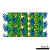 6188C 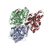 3j8yC M: map data used to model this data C: citing same article ( |
|---|---|
| Similar structure data |


-Links
 PDBj
PDBj


- Assembly
| Deposited unit | 
|
|---|---|
| 1 |
|
| Symmetry | Helical symmetry: (Circular symmetry: 1 / Dyad axis: no / N subunits divisor: 1 / Num. of operations: 1 / Rise per n subunits: 8.78 Å / Rotation per n subunits: -25.77 °) |
| Details | The reconstructed 14-protofilament microtubule is pseudo-symmetric, containing a seam with 3 starts per tubulin monomer, or 1.5 starts per tubulin dimer. |
-Components
| #1: Protein | Mass: 39238.145 Da / Num. of mol.: 1 Fragment: Truncated catalytic head domain (monomeric, UNP residues 1-349) Source method: isolated from a genetically manipulated source Source: (gene. exp.) Homo sapiens (human) / Gene: KIF5B, KNS, KNS1 / Production host:  |
|---|---|
| #2: Protein | Mass: 50204.445 Da / Num. of mol.: 1 / Source method: isolated from a natural source / Source: (natural) |
| #3: Protein | Mass: 49983.824 Da / Num. of mol.: 1 / Source method: isolated from a natural source / Source: (natural) |
| #4: Chemical | ChemComp-GTP /   Mass: 523.180 Da / Num. of mol.: 1 / Source method: obtained synthetically / Formula: C10H16N5O14P3 / Comment: GTP, energy-carrying molecule*YM Mass: 523.180 Da / Num. of mol.: 1 / Source method: obtained synthetically / Formula: C10H16N5O14P3 / Comment: GTP, energy-carrying molecule*YM |
| #5: Chemical | ChemComp-GDP /   Type: RNA linking / Mass: 443.201 Da / Num. of mol.: 1 / Source method: obtained synthetically / Formula: C10H15N5O11P2 / Comment: GDP, energy-carrying molecule*YM Type: RNA linking / Mass: 443.201 Da / Num. of mol.: 1 / Source method: obtained synthetically / Formula: C10H15N5O11P2 / Comment: GDP, energy-carrying molecule*YM |
-Experimental details
-Experiment
| Experiment | Method: ELECTRON MICROSCOPY |
|---|---|
| EM experiment | Aggregation state: FILAMENT / 3D reconstruction method: helical reconstruction |
- Sample preparation
| Component |
| |||||||||||||||||||||||||
|---|---|---|---|---|---|---|---|---|---|---|---|---|---|---|---|---|---|---|---|---|---|---|---|---|---|---|
| Molecular weight | Value: 0.135 MDa / Experimental value: NO | |||||||||||||||||||||||||
| Buffer solution | Name: 25 mM PIPES, 25 mM NaCl, 2 mM MgCl2, 1 mM EGTA / pH: 6.8 / Details: 25 mM PIPES, 25 mM NaCl, 2 mM MgCl2, 1 mM EGTA | |||||||||||||||||||||||||
| Specimen | Embedding applied: NO / Shadowing applied: NO / Staining applied: NO / Vitrification applied: YES | |||||||||||||||||||||||||
| Specimen support | Details: 300 mesh copper grid with homemade holey carbon | |||||||||||||||||||||||||
| Vitrification | Instrument: HOMEMADE PLUNGER / Cryogen name: ETHANE Details: No glow discharge was applied. After sample application to grid, liquid was mostly 'wicked' away by edgewise application of filter paper. Subsequently, blotting and plunge freezing were ...Details: No glow discharge was applied. After sample application to grid, liquid was mostly 'wicked' away by edgewise application of filter paper. Subsequently, blotting and plunge freezing were performed with ~0.5 second delay after blotting but prior to plunging into liquid ethane. Method: No glow discharge was applied. After sample application to grid, liquid was mostly 'wicked' away by edgewise application of filter paper. Subsequently, blotting and plunge freezing were ...Method: No glow discharge was applied. After sample application to grid, liquid was mostly 'wicked' away by edgewise application of filter paper. Subsequently, blotting and plunge freezing were performed with ~0.5 second delay after blotting but prior to plunging. |
- Electron microscopy imaging
| Experimental equipment |  Model: Tecnai F30 / Image courtesy: FEI Company |
|---|---|
| Microscopy | Model: FEI TECNAI F30 / Date: Apr 15, 2013 Details: 8K x 8K super-resolution mode was used. 10 frames total were collected. |
| Electron gun | Electron source:  FIELD EMISSION GUN / Accelerating voltage: 300 kV / Illumination mode: FLOOD BEAM FIELD EMISSION GUN / Accelerating voltage: 300 kV / Illumination mode: FLOOD BEAM |
| Electron lens | Mode: BRIGHT FIELD / Nominal magnification: 130000 X / Nominal defocus max: 2500 nm / Nominal defocus min: 1000 nm / Cs: 2 mm / Camera length: 0 mm |
| Specimen holder | Specimen holder model: GATAN LIQUID NITROGEN |
| Image recording | Electron dose: 15 e/Å2 / Film or detector model: GATAN K2 (4k x 4k) |
| Radiation | Protocol: SINGLE WAVELENGTH / Monochromatic (M) / Laue (L): M / Scattering type: x-ray |
| Radiation wavelength | Relative weight: 1 |
- Processing
| EM software |
| ||||||||||||
|---|---|---|---|---|---|---|---|---|---|---|---|---|---|
| CTF correction | Details: done within FREALIGN | ||||||||||||
| Helical symmerty | Angular rotation/subunit: 25.77 ° / Axial rise/subunit: 8.78 Å / Axial symmetry: C1 Details: The reconstructed 14-protofilament microtubule is pseudo-symmetric, containing a seam with 3 starts per tubulin monomer, or 1.5 starts per tubulin dimer. | ||||||||||||
| 3D reconstruction | Method: Single particle / Resolution: 5 Å / Resolution method: FSC 0.143 CUT-OFF / Num. of particles: 120783 / Nominal pixel size: 1.99 Å / Actual pixel size: 1.99 Å Details: Initial alignment was done using customized SPIDER scripts. Reconstruction and subsequent refinement were done by FREALIGN. Symmetry type: HELICAL | ||||||||||||
| Atomic model building | Protocol: FLEXIBLE FIT / Space: REAL Target criteria: RMSD from the starting structure was monitored for convergence. Details: REFINEMENT PROTOCOL--flexible DETAILS--MDFF was performed using explicit solvation. Side chains were excluded from the MDFF target potential. Following several equilibration steps, the ...Details: REFINEMENT PROTOCOL--flexible DETAILS--MDFF was performed using explicit solvation. Side chains were excluded from the MDFF target potential. Following several equilibration steps, the relative strength of the EM map potential (GSCALE term) was slowly increased from 0 to 1 over the course of 10 nanoseconds. The t = 1.2 ns time point was selected to represent the final fitted model, based on the approximate convergence of the RMSD from the starting structure. | ||||||||||||
| Atomic model building | 3D fitting-ID: 1 / Accession code: 4HNA / Initial refinement model-ID: 1 / PDB-ID: 4HNA / Source name: PDB / Type: experimental model
| ||||||||||||
| Refinement step | Cycle: LAST
|