 ムービー
ムービー コントローラー


+ データを開く

- 基本情報
基本情報
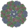
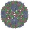
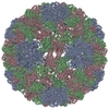
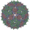
| 登録情報 | データベース: PDB / ID: 3j7n | ||||||
|---|---|---|---|---|---|---|---|
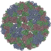
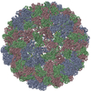
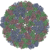
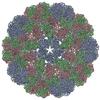
| タイトル | Virus model of brome mosaic virus (second half data set) | ||||||
 要素 要素 | Capsid protein | ||||||
 キーワード キーワード | VIRUS / capsid protein / BMV / beta barrel | ||||||
| 機能・相同性 |  機能・相同性情報 機能・相同性情報T=3 icosahedral viral capsid / host cell endoplasmic reticulum / viral nucleocapsid / ribonucleoprotein complex / structural molecule activity / RNA binding 類似検索 - 分子機能 | ||||||
| 生物種 |  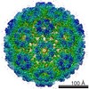 Brome mosaic virus (ウイルス) Brome mosaic virus (ウイルス) | ||||||
| 手法 | 電子顕微鏡法 / 単粒子再構成法 / クライオ電子顕微鏡法 / 解像度: 3.8 Å | ||||||
 データ登録者 データ登録者 | Wang, Z. / Hryc, C. / Bammes, B. / Afonine, P.V. / Jakana, J. / Chen, D.H. / Liu, X. / Baker, M.L. / Kao, C. / Ludtke, S.J. ...Wang, Z. / Hryc, C. / Bammes, B. / Afonine, P.V. / Jakana, J. / Chen, D.H. / Liu, X. / Baker, M.L. / Kao, C. / Ludtke, S.J. / Schmid, M.F. / Adams, P.D. / Chiu, W. | ||||||
 引用 引用 | ジャーナル: Nat Commun / 年: 2014 タイトル: An atomic model of brome mosaic virus using direct electron detection and real-space optimization. 著者: Zhao Wang / Corey F Hryc / Benjamin Bammes / Pavel V Afonine / Joanita Jakana / Dong-Hua Chen / Xiangan Liu / Matthew L Baker / Cheng Kao / Steven J Ludtke / Michael F Schmid / Paul D Adams / Wah Chiu /  要旨: Advances in electron cryo-microscopy have enabled structure determination of macromolecules at near-atomic resolution. However, structure determination, even using de novo methods, remains ...Advances in electron cryo-microscopy have enabled structure determination of macromolecules at near-atomic resolution. However, structure determination, even using de novo methods, remains susceptible to model bias and overfitting. Here we describe a complete workflow for data acquisition, image processing, all-atom modelling and validation of brome mosaic virus, an RNA virus. Data were collected with a direct electron detector in integrating mode and an exposure beyond the traditional radiation damage limit. The final density map has a resolution of 3.8 Å as assessed by two independent data sets and maps. We used the map to derive an all-atom model with a newly implemented real-space optimization protocol. The validity of the model was verified by its match with the density map and a previous model from X-ray crystallography, as well as the internal consistency of models from independent maps. This study demonstrates a practical approach to obtain a rigorously validated atomic resolution electron cryo-microscopy structure. | ||||||
| 履歴 |
|
- 構造の表示
構造の表示
| ムービー |
ムービービューア |
|---|---|
| 構造ビューア | 分子: MolmilJmol/JSmol |
- ダウンロードとリンク
-ダウンロード
| PDBx/mmCIF形式 | 3j7n.cif.gz | 102.1 KB | 表示 | PDBx/mmCIF形式 |
|---|---|---|---|---|
| PDB形式 | pdb3j7n.ent.gz | 80.5 KB | 表示 | PDB形式 |
| PDBx/mmJSON形式 | 3j7n.json.gz | ツリー表示 | PDBx/mmJSON形式 | |
| その他 |  その他のダウンロード その他のダウンロード |
-検証レポート
| アーカイブディレクトリ | https://data.pdbj.org/pub/pdb/validation_reports/j7/3j7nftp://data.pdbj.org/pub/pdb/validation_reports/j7/3j7n | HTTPS FTP |
|---|
-関連構造データ
| 関連構造データ | 6000MC 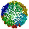 3j7lC 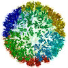 3j7mC M: このデータのモデリングに利用したマップデータ C: 同じ文献を引用 ( |
|---|---|
| 類似構造データ | |
| 電子顕微鏡画像生データ | EMPIAR-10010 (タイトル: Full virus map of Brome Mosaic Virus (micrographs and particle coordinates) Data size: 1.7 TB Data #1: Brome Mosaic Virus micrographs - non gain corrected [micrographs - multiframe] Data #2: Brome Mosaic Virus micrographs - gain corrected [micrographs - multiframe]) EMPIAR-10011 (タイトル: Full virus map of Brome Mosaic Virus (picked particles)Data size: 23.1 Data #1: Brome Mosaic Virus boxed particles [picked particles - single frame - processed]) |
-リンク
 PDBj
PDBj
- 集合体
集合体
| 登録構造単位 | 
|
|---|---|
| 1 | x 60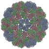
|
| 2 |
|
| 3 | x 5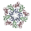
|
| 4 | x 6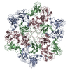
|
| 5 | 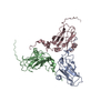
|
| 対称性 | 点対称性: (シェーンフリース記号: I (正20面体型対称)) |
-要素
| #1: タンパク質 | 分子量: 20411.525 Da / 分子数: 3 / 由来タイプ: 天然 / 由来: (天然) Brome mosaic virus (ウイルス) / 参照: UniProt: P03602 |
|---|
-実験情報
-実験
| 実験 | 手法: 電子顕微鏡法 |
|---|---|
| EM実験 | 試料の集合状態: PARTICLE / 3次元再構成法: 単粒子再構成法 |
- 試料調製
試料調製
| 構成要素 | 名称: Brome mosaic virus / タイプ: VIRUS |
|---|---|
| ウイルスについての詳細 | 中空か: NO / エンベロープを持つか: NO / ホストのカテゴリ: PLANTAE(HIGHER PLANTS) / 単離: STRAIN / タイプ: VIRION |
| 天然宿主 | 生物種: Triticum aestivum |
| 緩衝液 | pH: 5.2 |
| 試料 | 包埋: NO / シャドウイング: NO / 染色: NO / 凍結: YES |
| 急速凍結 | 装置: FEI VITROBOT MARK IV / 凍結剤: ETHANE / 湿度: 100 % / 凍結前の試料温度: 293 K / 詳細: Plunged into liquid ethane (FEI VITROBOT MARK IV). |
- 電子顕微鏡撮影
| 顕微鏡 | モデル: JEOL 3200FSC / 日付: 2013年1月10日 |
|---|---|
| 電子銃 | 電子線源:  FIELD EMISSION GUN / 加速電圧: 300 kV / 照射モード: FLOOD BEAM FIELD EMISSION GUN / 加速電圧: 300 kV / 照射モード: FLOOD BEAM |
| 電子レンズ | モード: BRIGHT FIELD / 倍率(公称値): 50000 X / 最大 デフォーカス(公称値): 2000 nm / 最小 デフォーカス(公称値): 500 nm / カメラ長: 0 mm |
| 試料ホルダ | 試料ホルダーモデル: JEOL 3200FSC CRYOHOLDER |
| 撮影 | フィルム・検出器のモデル: DIRECT ELECTRON DE-12 (4k x 3k) |
- 解析
解析
| EMソフトウェア |
| ||||||||||||
|---|---|---|---|---|---|---|---|---|---|---|---|---|---|
| 対称性 | 点対称性: I (正20面体型対称) | ||||||||||||
| 3次元再構成 | 手法: Single Particle / 解像度: 3.8 Å / 解像度の算出法: FSC 0.143 CUT-OFF / 粒子像の数: 30000 / ピクセルサイズ(公称値): 0.93 Å / ピクセルサイズ(実測値): 0.99 Å / 詳細: (Single particle--Applied symmetry: I) / 対称性のタイプ: POINT | ||||||||||||
| 精密化ステップ | サイクル: LAST
|
