 Movie
Movie Controller


+ Open data

- Basic information
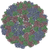
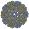
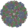
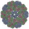
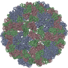
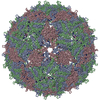
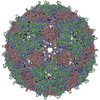
| Entry | Database: PDB / ID: 3j7l | ||||||
|---|---|---|---|---|---|---|---|
| Title | Full virus map of brome mosaic virus | ||||||
 Components Components | Capsid protein | ||||||
 Keywords Keywords | VIRUS / capsid protein / BMV / beta barrel | ||||||
| Function / homology |  Function and homology information Function and homology informationT=3 icosahedral viral capsid / host cell endoplasmic reticulum / viral nucleocapsid / ribonucleoprotein complex / structural molecule activity / RNA binding Similarity search - Function | ||||||
| Biological species |  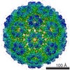 Brome mosaic virus Brome mosaic virus | ||||||
| Method | ELECTRON MICROSCOPY / single particle reconstruction / cryo EM / Resolution: 3.8 Å | ||||||
 Authors Authors | Wang, Z. / Hryc, C. / Bammes, B. / Afonine, P.V. / Jakana, J. / Chen, D.H. / Liu, X. / Baker, M.L. / Kao, C. / Ludtke, S.J. ...Wang, Z. / Hryc, C. / Bammes, B. / Afonine, P.V. / Jakana, J. / Chen, D.H. / Liu, X. / Baker, M.L. / Kao, C. / Ludtke, S.J. / Schmid, M.F. / Adams, P.D. / Chiu, W. | ||||||
 Citation Citation | Journal: Nat Commun / Year: 2014 Title: An atomic model of brome mosaic virus using direct electron detection and real-space optimization. Authors: Zhao Wang / Corey F Hryc / Benjamin Bammes / Pavel V Afonine / Joanita Jakana / Dong-Hua Chen / Xiangan Liu / Matthew L Baker / Cheng Kao / Steven J Ludtke / Michael F Schmid / Paul D Adams / Wah Chiu /  Abstract: Advances in electron cryo-microscopy have enabled structure determination of macromolecules at near-atomic resolution. However, structure determination, even using de novo methods, remains ...Advances in electron cryo-microscopy have enabled structure determination of macromolecules at near-atomic resolution. However, structure determination, even using de novo methods, remains susceptible to model bias and overfitting. Here we describe a complete workflow for data acquisition, image processing, all-atom modelling and validation of brome mosaic virus, an RNA virus. Data were collected with a direct electron detector in integrating mode and an exposure beyond the traditional radiation damage limit. The final density map has a resolution of 3.8 Å as assessed by two independent data sets and maps. We used the map to derive an all-atom model with a newly implemented real-space optimization protocol. The validity of the model was verified by its match with the density map and a previous model from X-ray crystallography, as well as the internal consistency of models from independent maps. This study demonstrates a practical approach to obtain a rigorously validated atomic resolution electron cryo-microscopy structure. | ||||||
| History |
|
- Structure visualization
| Movie |
Movie viewer |
|---|---|
| Structure viewer | Molecule: MolmilJmol/JSmol |
- Downloads & links
-Download
| PDBx/mmCIF format | 3j7l.cif.gz | 101.4 KB | Display | PDBx/mmCIF format |
|---|---|---|---|---|
| PDB format | pdb3j7l.ent.gz | 79.4 KB | Display | PDB format |
| PDBx/mmJSON format | 3j7l.json.gz | Tree view | PDBx/mmJSON format | |
| Others |  Other downloads Other downloads |
-Validation report
| Arichive directory | https://data.pdbj.org/pub/pdb/validation_reports/j7/3j7lftp://data.pdbj.org/pub/pdb/validation_reports/j7/3j7l | HTTPS FTP |
|---|
-Related structure data
| Related structure data | 6000MC 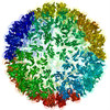 3j7mC 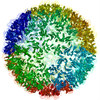 3j7nC M: map data used to model this data C: citing same article ( |
|---|---|
| Similar structure data | |
| EM raw data | EMPIAR-10010 (Title: Full virus map of Brome Mosaic Virus (micrographs and particle coordinates) Data size: 1.7 TB Data #1: Brome Mosaic Virus micrographs - non gain corrected [micrographs - multiframe] Data #2: Brome Mosaic Virus micrographs - gain corrected [micrographs - multiframe]) EMPIAR-10011 (Title: Full virus map of Brome Mosaic Virus (picked particles)Data size: 23.1 Data #1: Brome Mosaic Virus boxed particles [picked particles - single frame - processed]) |

-Links
 PDBj
PDBj
- Assembly
| Deposited unit | 
|
|---|---|
| 1 | x 60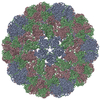
|
| 2 |
|
| 3 | x 5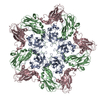
|
| 4 | x 6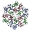
|
| 5 | 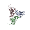
|
| Symmetry | Point symmetry: (Schoenflies symbol: I (icosahedral)) |
-Components
| #1: Protein | Mass: 20411.525 Da / Num. of mol.: 3 / Source method: isolated from a natural source / Source: (natural) Brome mosaic virus / References: UniProt: P03602 |
|---|
-Experimental details
-Experiment
| Experiment | Method: ELECTRON MICROSCOPY |
|---|---|
| EM experiment | Aggregation state: PARTICLE / 3D reconstruction method: single particle reconstruction |
- Sample preparation
| Component | Name: Brome mosaic virus / Type: VIRUS |
|---|---|
| Details of virus | Empty: NO / Enveloped: NO / Host category: PLANTAE(HIGHER PLANTS) / Isolate: STRAIN / Type: VIRION |
| Natural host | Organism: Triticum aestivum |
| Buffer solution | pH: 5.2 |
| Specimen | Embedding applied: NO / Shadowing applied: NO / Staining applied: NO / Vitrification applied: YES |
| Vitrification | Instrument: FEI VITROBOT MARK IV / Cryogen name: ETHANE / Humidity: 100 % / Chamber temperature: 293 K / Details: Plunged into liquid ethane (FEI VITROBOT MARK IV). |
- Electron microscopy imaging
| Microscopy | Model: JEOL 3200FSC / Date: Jan 10, 2013 |
|---|---|
| Electron gun | Electron source:  FIELD EMISSION GUN / Accelerating voltage: 300 kV / Illumination mode: FLOOD BEAM FIELD EMISSION GUN / Accelerating voltage: 300 kV / Illumination mode: FLOOD BEAM |
| Electron lens | Mode: BRIGHT FIELD / Nominal magnification: 50000 X / Nominal defocus max: 2000 nm / Nominal defocus min: 500 nm / Camera length: 0 mm |
| Specimen holder | Specimen holder model: JEOL 3200FSC CRYOHOLDER |
| Image recording | Film or detector model: DIRECT ELECTRON DE-12 (4k x 3k) |
- Processing
| EM software |
| ||||||||||||
|---|---|---|---|---|---|---|---|---|---|---|---|---|---|
| Symmetry | Point symmetry: I (icosahedral) | ||||||||||||
| 3D reconstruction | Method: Single Particle / Resolution: 3.8 Å / Resolution method: FSC 0.143 CUT-OFF / Num. of particles: 30000 / Nominal pixel size: 0.93 Å / Actual pixel size: 0.99 Å / Details: (Single particle--Applied symmetry: I) / Symmetry type: POINT | ||||||||||||
| Refinement step | Cycle: LAST
|
