 Movie
Movie Controller


+ Open data

- Basic information
| Entry | Database: PDB / ID: 3j6d | ||||||
|---|---|---|---|---|---|---|---|
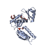
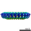
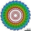
| Title | Model of the PrgH-PrgK periplasmic rings | ||||||
 Components Components |
| ||||||
 Keywords Keywords | STRUCTURAL PROTEIN / Bacterial secression macromolecular assemblies | ||||||
| Function / homology |  Function and homology information Function and homology information | ||||||
| Biological species |  Salmonella enterica subsp. enterica serovar Typhimurium str. LT2 (bacteria) Salmonella enterica subsp. enterica serovar Typhimurium str. LT2 (bacteria) | ||||||
| Method | ELECTRON MICROSCOPY / single particle reconstruction / cryo EM / Resolution: 11.7 Å | ||||||
 Authors Authors | Bergeron, J.R.C. / Strynadka, N.C.J. | ||||||
 Citation Citation | Journal: Science / Year: 2011 Title: Three-dimensional model of Salmonella's needle complex at subnanometer resolution. Authors: Oliver Schraidt / Thomas C Marlovits /  Abstract: Type III secretion systems (T3SSs) are essential virulence factors used by many Gram-negative bacteria to inject proteins that make eukaryotic host cells accessible to invasion. The T3SS core ...Type III secretion systems (T3SSs) are essential virulence factors used by many Gram-negative bacteria to inject proteins that make eukaryotic host cells accessible to invasion. The T3SS core structure, the needle complex (NC), is a ~3.5 megadalton-sized, oligomeric, membrane-embedded complex. Analyzing cryo-electron microscopy images of top views of NCs or NC substructures from Salmonella typhimurium revealed a 24-fold symmetry for the inner rings and a 15-fold symmetry for the outer rings, giving an overall C3 symmetry. Local refinement and averaging showed the organization of the central core and allowed us to reconstruct a subnanometer composite structure of the NC, which together with confident docking of atomic structures reveal insights into its overall organization and structural requirements during assembly. | ||||||
| History |
| ||||||
| Remark 0 | THIS ENTRY 3J6D CONTAINS A STRUCTURAL MODEL FIT TO AN ELECTRON MICROSCOPY MAP (EMD-1875) DETERMINED ...THIS ENTRY 3J6D CONTAINS A STRUCTURAL MODEL FIT TO AN ELECTRON MICROSCOPY MAP (EMD-1875) DETERMINED ORIGINALLY BY AUTHORS: O.SCHRAIDT, T.C.MARLOVITS |
- Structure visualization
| Movie |
Movie viewer |
|---|---|
| Structure viewer | Molecule: MolmilJmol/JSmol |
- Downloads & links
-Download
| PDBx/mmCIF format | 3j6d.cif.gz | 1.4 MB | Display | PDBx/mmCIF format |
|---|---|---|---|---|
| PDB format | pdb3j6d.ent.gz | 1.1 MB | Display | PDB format |
| PDBx/mmJSON format | 3j6d.json.gz | Tree view | PDBx/mmJSON format | |
| Others |  Other downloads Other downloads |
-Validation report
| Arichive directory | https://data.pdbj.org/pub/pdb/validation_reports/j6/3j6dftp://data.pdbj.org/pub/pdb/validation_reports/j6/3j6d | HTTPS FTP |
|---|
-Related structure data
| Related structure data | 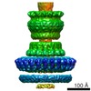 1875M  2mkyC 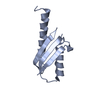 4oycC 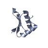 4w4mC C: citing same article ( M: map data used to model this data |
|---|---|
| Similar structure data |


-Links
 PDBj
PDBj


- Assembly
| Deposited unit | 
|
|---|---|
| 1 |
|
-Components
| #1: Protein | Mass: 44509.367 Da / Num. of mol.: 24 Source method: isolated from a genetically manipulated source Source: (gene. exp.) Salmonella enterica subsp. enterica serovar Typhimurium str. LT2 (bacteria)Gene: prgH, STM2874 / Production host: #2: Protein | Mass: 28245.287 Da / Num. of mol.: 24 Source method: isolated from a genetically manipulated source Source: (gene. exp.) Salmonella enterica subsp. enterica serovar Typhimurium str. LT2 (bacteria)Gene: SEETLT22_01090 / Production host: |
|---|
-Experimental details
-Experiment
| Experiment | Method: ELECTRON MICROSCOPY |
|---|---|
| EM experiment | Aggregation state: PARTICLE / 3D reconstruction method: single particle reconstruction |
- Sample preparation
| Component | Name: PrgH-PrgK periplasmic rings / Type: COMPLEX |
|---|---|
| Buffer solution | pH: 7.5 |
| Specimen | Embedding applied: NO / Shadowing applied: NO / Staining applied: NO / Vitrification applied: YES |
| Vitrification | Cryogen name: ETHANE |
- Electron microscopy imaging
| Experimental equipment |  Model: Tecnai Polara / Image courtesy: FEI Company |
|---|---|
| Microscopy | Model: FEI POLARA 300 |
| Electron gun | Electron source:  FIELD EMISSION GUN / Accelerating voltage: 300 kV / Illumination mode: FLOOD BEAM FIELD EMISSION GUN / Accelerating voltage: 300 kV / Illumination mode: FLOOD BEAM |
| Electron lens | Mode: BRIGHT FIELD / Nominal magnification: 93000 X |
| Image recording | Film or detector model: GENERIC CCD |
- Processing
| EM software |
| ||||||||||||||||||||||||||||
|---|---|---|---|---|---|---|---|---|---|---|---|---|---|---|---|---|---|---|---|---|---|---|---|---|---|---|---|---|---|
| Symmetry | Point symmetry: C24 (24 fold cyclic) | ||||||||||||||||||||||||||||
| 3D reconstruction | Resolution: 11.7 Å / Resolution method: FSC 0.5 CUT-OFF / Num. of particles: 37171 / Symmetry type: POINT | ||||||||||||||||||||||||||||
| Atomic model building | Protocol: OTHER / Space: REAL / Details: METHOD--EM-guided symmetrical modeling | ||||||||||||||||||||||||||||
| Atomic model building |
| ||||||||||||||||||||||||||||
| Refinement step | Cycle: LAST
|

