Movie
Movie Controller

[English] 日本語
 Yorodumi
- EMDB-12193: Salmonella flagellar basal body assembly intermediate - P ring al... -
+ Open data

- Basic information
| Entry | Database: EMDB / ID: EMD-12193 | |||||||||||||||
|---|---|---|---|---|---|---|---|---|---|---|---|---|---|---|---|---|
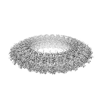
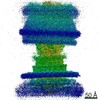
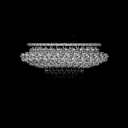
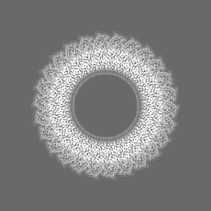
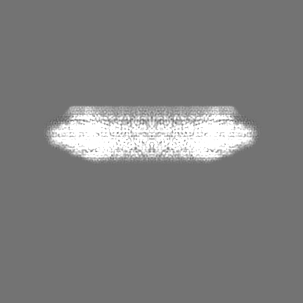
| Title | Salmonella flagellar basal body assembly intermediate - P ring alone structure | |||||||||||||||
 Map data Map data | refinement volume | |||||||||||||||
 Sample Sample |
| |||||||||||||||
 Keywords Keywords | bacterial flagellum rod bacterial flagellum export gate basal body / PROTEIN TRANSPORT | |||||||||||||||
| Function / homology | bacterial-type flagellum basal body, distal rod, P ring / Flagellar P-ring protein / Flagellar P-ring protein / bacterial-type flagellum-dependent cell motility / outer membrane-bounded periplasmic space / structural molecule activity / Flagellar P-ring protein Function and homology information Function and homology information | |||||||||||||||
| Biological species |  Salmonella enterica subsp. enterica serovar Typhi (bacteria) / Salmonella typhimurium (strain LT2 / SGSC1412 / ATCC 700720) (bacteria) Salmonella enterica subsp. enterica serovar Typhi (bacteria) / Salmonella typhimurium (strain LT2 / SGSC1412 / ATCC 700720) (bacteria) | |||||||||||||||
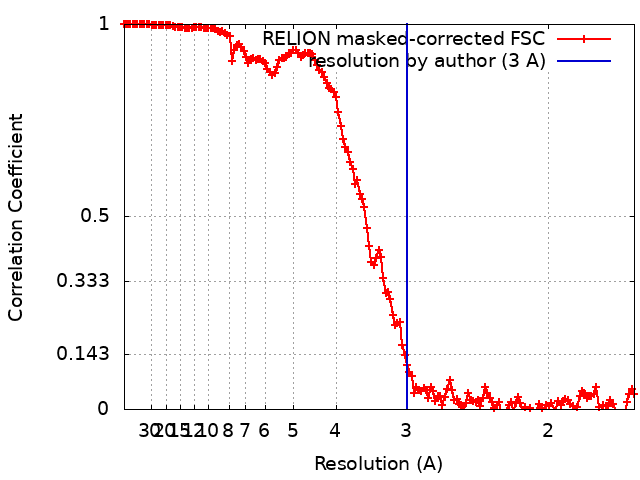
| Method | single particle reconstruction / cryo EM / Resolution: 3.0 Å | |||||||||||||||
 Authors Authors | Johnson S / Furlong E | |||||||||||||||
| Funding support |  United Kingdom, 4 items United Kingdom, 4 items
| |||||||||||||||
 Citation Citation | Journal: Nat Microbiol / Year: 2021 Title: Molecular structure of the intact bacterial flagellar basal body. Authors: Steven Johnson / Emily J Furlong / Justin C Deme / Ashley L Nord / Joseph J E Caesar / Fabienne F V Chevance / Richard M Berry / Kelly T Hughes / Susan M Lea /   Abstract: The bacterial flagellum is a macromolecular protein complex that enables motility in many species. Bacterial flagella self-assemble a strong, multicomponent drive shaft that couples rotation in the ...The bacterial flagellum is a macromolecular protein complex that enables motility in many species. Bacterial flagella self-assemble a strong, multicomponent drive shaft that couples rotation in the inner membrane to the micrometre-long flagellar filament that powers bacterial swimming in viscous fluids. Here, we present structures of the intact Salmonella flagellar basal body, encompassing the inner membrane rotor, drive shaft and outer-membrane bushing, solved using cryo-electron microscopy to resolutions of 2.2-3.7 Å. The structures reveal molecular details of how 173 protein molecules of 13 different types assemble into a complex spanning two membranes and a cell wall. The helical drive shaft at one end is intricately interwoven with the rotor component with both the export gate complex and the proximal rod forming interactions with the MS-ring. At the other end, the drive shaft distal rod passes through the LP-ring bushing complex, which functions as a molecular bearing anchored in the outer membrane through interactions with the lipopolysaccharide. The in situ structure of a protein complex capping the drive shaft provides molecular insights into the assembly process of this molecular machine. | |||||||||||||||
| History |
|
- Structure visualization
| Movie |
Movie viewer |
|---|---|
| Structure viewer | EM map: SurfViewMolmilJmol/JSmol |
| Supplemental images |
- Downloads & links
-EMDB archive
| Map data | emd_12193.map.gz | 241.6 MB | EMDB map data format | |
|---|---|---|---|---|
| Header (meta data) | emd-12193-v30.xmlemd-12193.xml | 16.8 KB 16.8 KB | Display Display | EMDB header |
| FSC (resolution estimation) | emd_12193_fsc.xml | 15.3 KB | Display | FSC data file |
| Images |  emd_12193.png emd_12193.png | 39.8 KB | ||
| Filedesc metadata | emd-12193.cif.gz | 5.1 KB | ||
| Others | emd_12193_additional_1.map.gzemd_12193_half_map_1.map.gzemd_12193_half_map_2.map.gz | 25.7 MB 242.8 MB 242.8 MB | ||
| Archive directory |  http://ftp.pdbj.org/pub/emdb/structures/EMD-12193ftp://ftp.pdbj.org/pub/emdb/structures/EMD-12193 http://ftp.pdbj.org/pub/emdb/structures/EMD-12193ftp://ftp.pdbj.org/pub/emdb/structures/EMD-12193 | HTTPS FTP |
-Related structure data
| Related structure data |  7bj2MC  7bglC 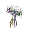 7bhqC  7binC  7bk0C  7nvgC M: atomic model generated by this map C: citing same article ( |
|---|---|
| Similar structure data |

-Links
| EMDB pages | EMDB (EBI/PDBe) / EMDataResource |
|---|---|
| Related items in Molecule of the Month |

-Map
| File | Download / File: emd_12193.map.gz / Format: CCP4 / Size: 307.5 MB / Type: IMAGE STORED AS FLOATING POINT NUMBER (4 BYTES) | ||||||||||||||||||||||||||||||||||||||||||||||||||||||||||||
|---|---|---|---|---|---|---|---|---|---|---|---|---|---|---|---|---|---|---|---|---|---|---|---|---|---|---|---|---|---|---|---|---|---|---|---|---|---|---|---|---|---|---|---|---|---|---|---|---|---|---|---|---|---|---|---|---|---|---|---|---|---|
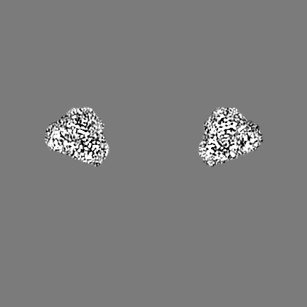
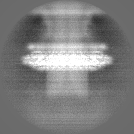
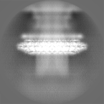
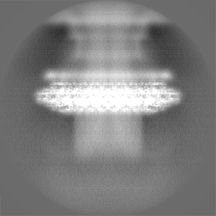
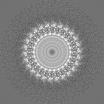
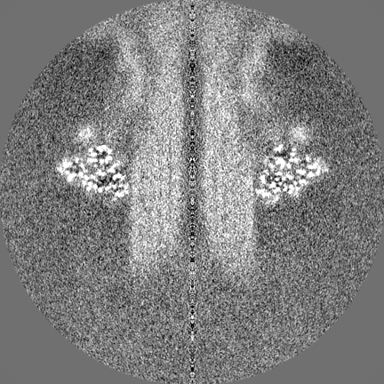
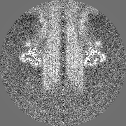
| Annotation | refinement volume | ||||||||||||||||||||||||||||||||||||||||||||||||||||||||||||
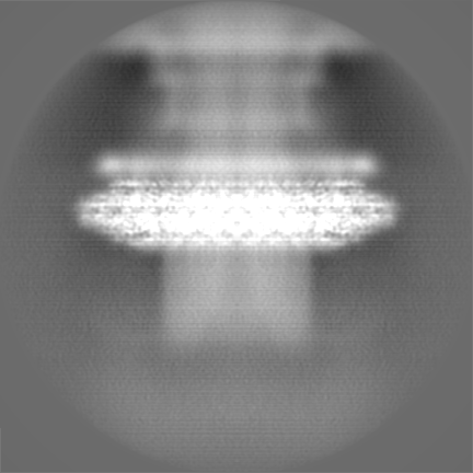
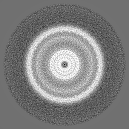
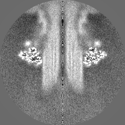
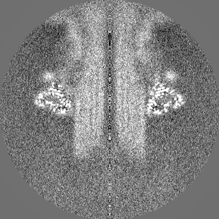
| Projections & slices | Image control
Images are generated by Spider. | ||||||||||||||||||||||||||||||||||||||||||||||||||||||||||||
| Voxel size | X=Y=Z: 0.832 Å | ||||||||||||||||||||||||||||||||||||||||||||||||||||||||||||
| Density |
| ||||||||||||||||||||||||||||||||||||||||||||||||||||||||||||
| Symmetry | Space group: 1 | ||||||||||||||||||||||||||||||||||||||||||||||||||||||||||||
| Details | EMDB XML:
CCP4 map header:
| ||||||||||||||||||||||||||||||||||||||||||||||||||||||||||||
 Z (Sec.)
 Y (Row.)
 X (Col.)


-Supplemental data
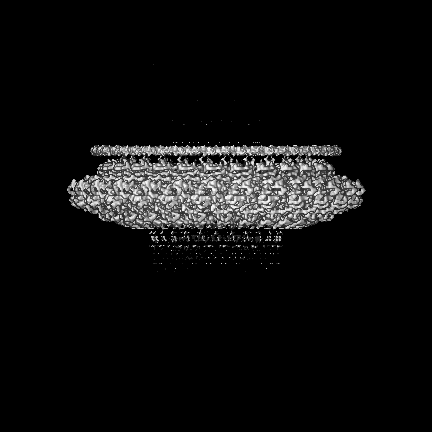
-Additional map: post processed volume
| File | emd_12193_additional_1.map | ||||||||||||
|---|---|---|---|---|---|---|---|---|---|---|---|---|---|
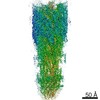
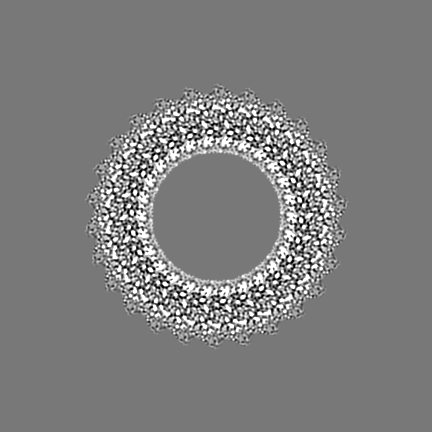
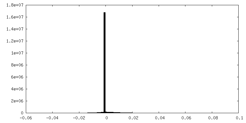
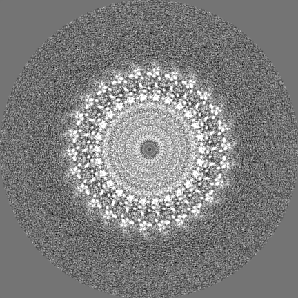
| Annotation | post processed volume | ||||||||||||
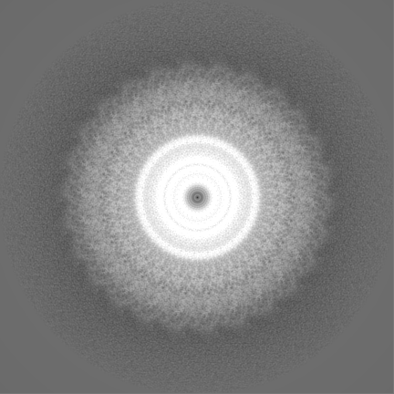
| Projections & Slices |
| ||||||||||||
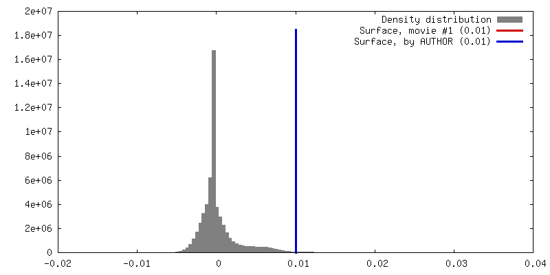
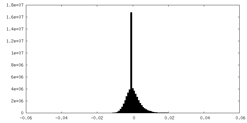
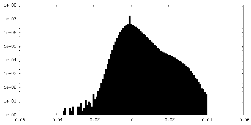
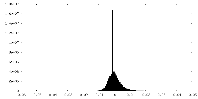
| Density Histograms |

-Half map: half map 1
| File | emd_12193_half_map_1.map | ||||||||||||
|---|---|---|---|---|---|---|---|---|---|---|---|---|---|
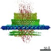
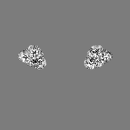
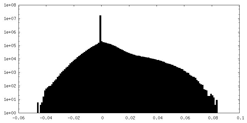
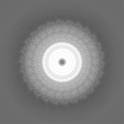
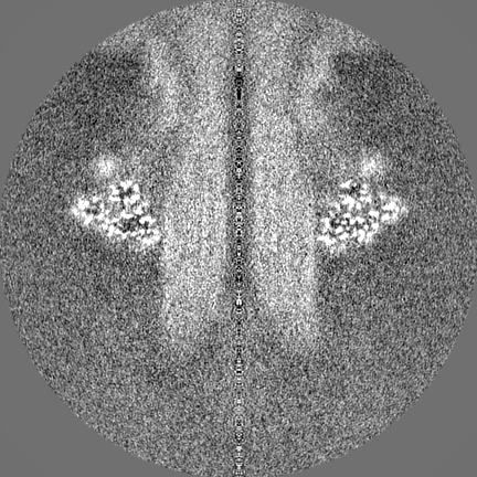
| Annotation | half map 1 | ||||||||||||
| Projections & Slices |
| ||||||||||||
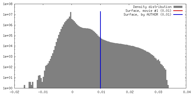
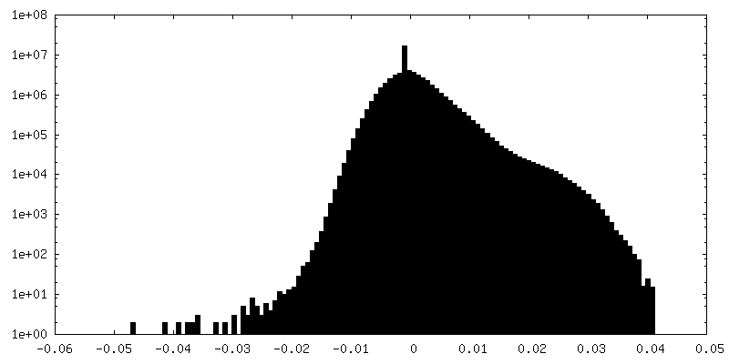
| Density Histograms |
-Half map: half map 2
| File | emd_12193_half_map_2.map | ||||||||||||
|---|---|---|---|---|---|---|---|---|---|---|---|---|---|
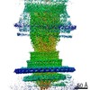
| Annotation | half map 2 | ||||||||||||
| Projections & Slices |
| ||||||||||||
| Density Histograms |

- Sample components
-Entire : Flagellar P ring assembly intermediate
| Entire | Name: Flagellar P ring assembly intermediate |
|---|---|
| Components |
|
-Supramolecule #1: Flagellar P ring assembly intermediate
| Supramolecule | Name: Flagellar P ring assembly intermediate / type: complex / ID: 1 / Parent: 0 / Macromolecule list: all |
|---|---|
| Source (natural) | Organism: Salmonella enterica subsp. enterica serovar Typhi (bacteria) |
-Macromolecule #1: Flagellar P-ring protein
| Macromolecule | Name: Flagellar P-ring protein / type: protein_or_peptide / ID: 1 / Number of copies: 26 / Enantiomer: LEVO |
|---|---|
| Source (natural) | Organism: Salmonella typhimurium (strain LT2 / SGSC1412 / ATCC 700720) (bacteria) Strain: LT2 / SGSC1412 / ATCC 700720 |
| Molecular weight | Theoretical: 38.194176 KDa |
| Sequence | String: MFKALAGIVL ALVATLAHAE RIRDLTSVQG VRENSLIGYG LVVGLDGTGD QTTQTPFTTQ TLNNMLSQLG ITVPTGTNMQ LKNVAAVMV TASYPPFARQ GQTIDVVVSS MGNAKSLRGG TLLMTPLKGV DSQVYALAQG NILVGGAGAS AGGSSVQVNQ L NGGRITNG ...String: MFKALAGIVL ALVATLAHAE RIRDLTSVQG VRENSLIGYG LVVGLDGTGD QTTQTPFTTQ TLNNMLSQLG ITVPTGTNMQ LKNVAAVMV TASYPPFARQ GQTIDVVVSS MGNAKSLRGG TLLMTPLKGV DSQVYALAQG NILVGGAGAS AGGSSVQVNQ L NGGRITNG AIIERELPTQ FGAGNTINLQ LNDEDFTMAQ QITDAINRAR GYGSATALDA RTVQVRVPSG NSSQVRFLAD IQ NMEVNVT PQDAKVVINS RTGSVVMNRE VTLDSCAVAQ GNLSVTVNRQ LNVNQPNTPF GGGQTVVTPQ TQIDLRQSGG SLQ SVRSSA NLNSVVRALN ALGATPMDLM SILQSMQSAG CLRAKLEII UniProtKB: Flagellar P-ring protein |
-Experimental details
-Structure determination
| Method | cryo EM |
|---|---|
 Processing Processing | single particle reconstruction |
| Aggregation state | particle |
-Sample preparation
| Buffer | pH: 8 |
|---|---|
| Vitrification | Cryogen name: ETHANE |
- Electron microscopy
| Microscope | FEI TITAN KRIOS |
|---|---|
| Image recording | Film or detector model: GATAN K3 BIOQUANTUM (6k x 4k) / Average electron dose: 59.0 e/Å2 |
| Electron beam | Acceleration voltage: 300 kV / Electron source:  FIELD EMISSION GUN FIELD EMISSION GUN |
| Electron optics | Illumination mode: FLOOD BEAM / Imaging mode: BRIGHT FIELD |
| Experimental equipment |  Model: Titan Krios / Image courtesy: FEI Company |