Movie
Movie Controller

[English] 日本語
 Yorodumi
- PDB-3iyd: Three-dimensional EM structure of an intact activator-dependent t... -
+ Open data

- Basic information
| Entry | Database: PDB / ID: 3iyd | ||||||
|---|---|---|---|---|---|---|---|
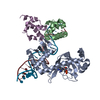
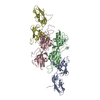
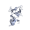
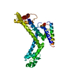
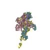
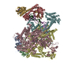
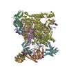
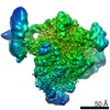
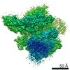
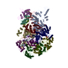
| Title | Three-dimensional EM structure of an intact activator-dependent transcription initiation complex | ||||||
 Components Components |
| ||||||
 Keywords Keywords | TRANSCRIPTION/DNA / transcription / initiation / Class I / activator / RNA polymerase / holoenzyme / sigma70 / open complex / CAP / CRP / cAMP-dependent / DNA / prokaryotic / DNA-directed RNA polymerase / Nucleotidyltransferase / Transferase / DNA-binding / Sigma factor / Transcription regulation / cAMP / cAMP-binding / Nucleotide-binding / TRANSCRIPTION-DNA COMPLEX | ||||||
| Function / homology |  Function and homology information Function and homology informationcarbon catabolite repression of transcription / sigma factor antagonist complex / DNA binding, bending / RNA polymerase complex / submerged biofilm formation / cellular response to cell envelope stress / minor groove of adenine-thymine-rich DNA binding / regulation of DNA-templated transcription initiation / sigma factor activity / bacterial-type flagellum assembly ...carbon catabolite repression of transcription / sigma factor antagonist complex / DNA binding, bending / RNA polymerase complex / submerged biofilm formation / cellular response to cell envelope stress / minor groove of adenine-thymine-rich DNA binding / regulation of DNA-templated transcription initiation / sigma factor activity / bacterial-type flagellum assembly / bacterial-type RNA polymerase core enzyme binding / cytosolic DNA-directed RNA polymerase complex / bacterial-type flagellum-dependent cell motility / nitrate assimilation / cAMP binding / regulation of DNA-templated transcription elongation / transcription elongation factor complex / DNA-directed RNA polymerase complex / transcription antitermination / cell motility / DNA-templated transcription initiation / protein-DNA complex / ribonucleoside binding / DNA-directed RNA polymerase / DNA-directed RNA polymerase activity / response to heat / protein-containing complex assembly / sequence-specific DNA binding / intracellular iron ion homeostasis / protein dimerization activity / transcription cis-regulatory region binding / DNA-binding transcription factor activity / response to antibiotic / negative regulation of DNA-templated transcription / regulation of DNA-templated transcription / positive regulation of DNA-templated transcription / DNA-templated transcription / magnesium ion binding / DNA binding / zinc ion binding / membrane / identical protein binding / cytoplasm / cytosol Similarity search - Function | ||||||
| Biological species |  | ||||||
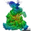
| Method | ELECTRON MICROSCOPY / single particle reconstruction / negative staining / Resolution: 19.8 Å | ||||||
 Authors Authors | Hudson, B.P. / Quispe, J. / Lara, S. / Kim, Y. / Berman, H. / Arnold, E. / Ebright, R.H. / Lawson, C.L. | ||||||
 Citation Citation | Journal: Proc Natl Acad Sci U S A / Year: 2009 Title: Three-dimensional EM structure of an intact activator-dependent transcription initiation complex. Authors: Brian P Hudson / Joel Quispe / Samuel Lara-González / Younggyu Kim / Helen M Berman / Eddy Arnold / Richard H Ebright / Catherine L Lawson /  Abstract: We present the experimentally determined 3D structure of an intact activator-dependent transcription initiation complex comprising the Escherichia coli catabolite activator protein (CAP), RNA ...We present the experimentally determined 3D structure of an intact activator-dependent transcription initiation complex comprising the Escherichia coli catabolite activator protein (CAP), RNA polymerase holoenzyme (RNAP), and a DNA fragment containing positions -78 to +20 of a Class I CAP-dependent promoter with a CAP site at position -61.5 and a premelted transcription bubble. A 20-A electron microscopy reconstruction was obtained by iterative projection-based matching of single particles visualized in carbon-sandwich negative stain and was fitted using atomic coordinate sets for CAP, RNAP, and DNA. The structure defines the organization of a Class I CAP-RNAP-promoter complex and supports previously proposed interactions of CAP with RNAP alpha subunit C-terminal domain (alphaCTD), interactions of alphaCTD with sigma(70) region 4, interactions of CAP and RNAP with promoter DNA, and phased-DNA-bend-dependent partial wrapping of DNA around the complex. The structure also reveals the positions and shapes of species-specific domains within the RNAP beta', beta, and sigma(70) subunits. | ||||||
| History |
| ||||||
| Remark 650 | HELIX DETERMINATION METHOD: AUTHOR DETERMINED | ||||||
| Remark 700 | SHEET DETERMINATION METHOD: AUTHOR DETERMINED |
- Structure visualization
| Movie |
Movie viewer |
|---|---|
| Structure viewer | Molecule: MolmilJmol/JSmol |
- Downloads & links
-Download
| PDBx/mmCIF format | 3iyd.cif.gz | 778.6 KB | Display | PDBx/mmCIF format |
|---|---|---|---|---|
| PDB format | pdb3iyd.ent.gz | 598.4 KB | Display | PDB format |
| PDBx/mmJSON format | 3iyd.json.gz | Tree view | PDBx/mmJSON format | |
| Others |  Other downloads Other downloads |
-Validation report
| Arichive directory | https://data.pdbj.org/pub/pdb/validation_reports/iy/3iydftp://data.pdbj.org/pub/pdb/validation_reports/iy/3iyd | HTTPS FTP |
|---|
-Related structure data
| Related structure data | 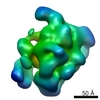 5127MC M: map data used to model this data C: citing same article ( |
|---|---|
| Similar structure data |

-Links
 PDBj
PDBj


- Assembly
| Deposited unit | 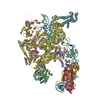
|
|---|---|
| 1 |
|
-Components
-DNA-directed RNA polymerase subunit ... , 4 types, 5 molecules ABCDE
| #1: Protein | Mass: 36558.680 Da / Num. of mol.: 2 Source method: isolated from a genetically manipulated source Source: (gene. exp.) Gene: b3295, JW3257, pez, phs, rpoA, RpoB, RpoC, RpoD, RpoZ, sez Plasmid: pEcABC-H6, pRSFduet-sigma, pCDF-omega / Production host: #2: Protein | | Mass: 150820.875 Da / Num. of mol.: 1 Source method: isolated from a genetically manipulated source Source: (gene. exp.) Gene: rpoB, groN, nitB, rif, ron, stl, stv, tabD, b3987, JW3950 Production host: #3: Protein | | Mass: 156195.625 Da / Num. of mol.: 1 / Source method: obtained synthetically / Details: Purchased from IDT and annealed / References: UniProt: P0A8T7, DNA-directed RNA polymerase #4: Protein | | Mass: 10118.352 Da / Num. of mol.: 1 Source method: isolated from a genetically manipulated source Source: (gene. exp.) |
|---|
-Protein , 2 types, 3 molecules FGH
| #5: Protein | Mass: 70326.234 Da / Num. of mol.: 1 Source method: isolated from a genetically manipulated source Source: (gene. exp.) |
|---|---|
| #6: Protein | Mass: 23541.242 Da / Num. of mol.: 2 Source method: isolated from a genetically manipulated source Source: (gene. exp.) |
-DNA chain , 2 types, 2 molecules IJ
| #7: DNA chain | Mass: 30220.389 Da / Num. of mol.: 1 / Source method: obtained synthetically |
|---|---|
| #8: DNA chain | Mass: 30097.295 Da / Num. of mol.: 1 / Source method: obtained synthetically |
-Non-polymers , 1 types, 2 molecules 
| #9: Chemical |  Mass: 329.206 Da / Num. of mol.: 2 / Source method: obtained synthetically / Formula: C10H12N5O6P Mass: 329.206 Da / Num. of mol.: 2 / Source method: obtained synthetically / Formula: C10H12N5O6P |
|---|
-Experimental details
-Experiment
| Experiment | Method: ELECTRON MICROSCOPY |
|---|---|
| EM experiment | Aggregation state: PARTICLE / 3D reconstruction method: single particle reconstruction |
- Sample preparation
| Component |
| |||||||||||||||||||||||||
|---|---|---|---|---|---|---|---|---|---|---|---|---|---|---|---|---|---|---|---|---|---|---|---|---|---|---|
| Molecular weight | Value: 0.57 MDa / Experimental value: NO | |||||||||||||||||||||||||
| Buffer solution | Name: 25 mM HEPES, 100 mM KCl, 10 mM MgCl2, 1 mM DTT, 0.2 mM cAMP pH: 8 Details: 25 mM HEPES, 100 mM KCl, 10 mM MgCl2, 1 mM DTT, 0.2 mM cAMP | |||||||||||||||||||||||||
| Specimen | Conc.: 6.18 mg/ml / Embedding applied: NO / Shadowing applied: NO / Staining applied: YES / Vitrification applied: NO Details: 25mM HEPES, 100mM KCl, 10mM MgCl2, 1mM DTT, 0.2mM cAMP | |||||||||||||||||||||||||
| EM staining | Type: NEGATIVE / Material: Uranyl Formate | |||||||||||||||||||||||||
| Specimen support | Details: 400-mesh copper 2.0x0.5 hole pattern C-flat grids (Protochips, Inc.) with a thin layer of continuous carbon floated on top |
- Electron microscopy imaging
| Experimental equipment |  Model: Tecnai F20 / Image courtesy: FEI Company |
|---|---|
| Microscopy | Model: FEI TECNAI F20 / Date: Nov 4, 2008 / Details: 15 um pixel size on detector |
| Electron gun | Electron source:  FIELD EMISSION GUN / Accelerating voltage: 120 kV / Illumination mode: FLOOD BEAM FIELD EMISSION GUN / Accelerating voltage: 120 kV / Illumination mode: FLOOD BEAM |
| Electron lens | Mode: BRIGHT FIELD / Nominal magnification: 50000 X / Nominal defocus max: 1500 nm / Nominal defocus min: 500 nm / Cs: 2 mm / Camera length: 0 mm |
| Specimen holder | Specimen holder model: SIDE ENTRY, EUCENTRIC Specimen holder type: standard side-entry room-temperature stage Temperature: 293 K / Tilt angle max: 0 ° / Tilt angle min: 0 ° |
| Image recording | Electron dose: 16 e/Å2 / Film or detector model: TVIPS TEMCAM-F415 (4k x 4k) |
| Radiation | Protocol: SINGLE WAVELENGTH / Monochromatic (M) / Laue (L): M / Scattering type: x-ray |
| Radiation wavelength | Relative weight: 1 |
- Processing
| EM software |
| ||||||||||||||||||||||||||||||
|---|---|---|---|---|---|---|---|---|---|---|---|---|---|---|---|---|---|---|---|---|---|---|---|---|---|---|---|---|---|---|---|
| CTF correction | Details: ACE | ||||||||||||||||||||||||||||||
| Symmetry | Point symmetry: C1 (asymmetric) | ||||||||||||||||||||||||||||||
| 3D reconstruction | Method: projection matching / Resolution: 19.8 Å / Resolution method: FSC 0.5 CUT-OFF / Num. of particles: 14097 Details: EMAN interleaved with SPIDER correspondence analysis ( Details about the particle: 32816 particles were automatically selected by the Appion DoGpicker initially ) Num. of class averages: 280 / Symmetry type: POINT | ||||||||||||||||||||||||||||||
| Atomic model building | Protocol: FLEXIBLE FIT / Space: REAL / Target criteria: map-derived potential energy Details: REFINEMENT PROTOCOL--rigid body, Yup.scx simulated annealing DETAILS--A complete ternary complex model was generated using multiple PDB entries, with a homology modelling step for RNAP. The ...Details: REFINEMENT PROTOCOL--rigid body, Yup.scx simulated annealing DETAILS--A complete ternary complex model was generated using multiple PDB entries, with a homology modelling step for RNAP. The model was regularized with PHENIX and the refined against the EM map with Yup.scx using default parameters. | ||||||||||||||||||||||||||||||
| Atomic model building | 3D fitting-ID: 1 / Source name: PDB / Type: experimental model
| ||||||||||||||||||||||||||||||
| Refinement step | Cycle: LAST
|