 Movie
Movie Controller

[English] 日本語
 Yorodumi
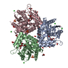
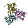
- PDB-3ix2: CRYSTAL STRUCTURE OF PURINE NUCLEOSIDE PHOSPHORYLASE FROM MYCOBAC... -
+ Open data

- Basic information
| Entry | Database: PDB / ID: 3ix2 | ||||||
|---|---|---|---|---|---|---|---|
| Title | CRYSTAL STRUCTURE OF PURINE NUCLEOSIDE PHOSPHORYLASE FROM MYCOBACTERIUM TUBERCULOSIS IN COMPLEX WITH ACYCLOVIR | ||||||
 Components Components | Purine nucleoside phosphorylase | ||||||
 Keywords Keywords | TRANSFERASE / Mycobacterium tuberculosis / Purine Nucleoside Phosphorylase / Acyclovir | ||||||
| Function / homology |  Function and homology information Function and homology informationnucleoside metabolic process / purine-nucleoside phosphorylase / purine-nucleoside phosphorylase activity / cytoplasm Similarity search - Function | ||||||
| Biological species |  Mycobacterium tuberculosis variant bovis AF2122/97 (bacteria) Mycobacterium tuberculosis variant bovis AF2122/97 (bacteria) | ||||||
| Method |  X-RAY DIFFRACTION / SYNCHROTRON / MOLECULAR REPLACEMENT / Resolution: 2.1 Å X-RAY DIFFRACTION / SYNCHROTRON / MOLECULAR REPLACEMENT / Resolution: 2.1 Å | ||||||
 Authors Authors | de Azevedo Jr., W.F. / Basso, L.A. / Santos, D.S. | ||||||
 Citation Citation | Journal: Biochimie / Year: 2012 Title: Crystal structure and molecular dynamics studies of purine nucleoside phosphorylase from Mycobacterium tuberculosis associated with acyclovir. Authors: Caceres, R.A. / Timmers, L.F. / Ducati, R.G. / da Silva, D.O. / Basso, L.A. / de Azevedo Jr., W.F. / Santos, D.S. | ||||||
| History |
|
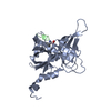
- Structure visualization
| Structure viewer | Molecule: MolmilJmol/JSmol |
|---|
- Downloads & links
-Download
| PDBx/mmCIF format | 3ix2.cif.gz | 160.9 KB | Display | PDBx/mmCIF format |
|---|---|---|---|---|
| PDB format | pdb3ix2.ent.gz | 127.5 KB | Display | PDB format |
| PDBx/mmJSON format | 3ix2.json.gz | Tree view | PDBx/mmJSON format | |
| Others |  Other downloads Other downloads |
-Validation report
| Arichive directory | https://data.pdbj.org/pub/pdb/validation_reports/ix/3ix2ftp://data.pdbj.org/pub/pdb/validation_reports/ix/3ix2 | HTTPS FTP |
|---|
-Related structure data
| Related structure data | 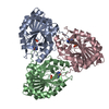 1n3iS S: Starting model for refinement |
|---|---|
| Similar structure data |


-Links
 PDBj
PDBj- Assembly
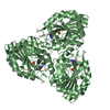
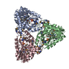
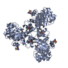
| Deposited unit | 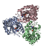
| ||||||||
|---|---|---|---|---|---|---|---|---|---|
| 1 |
| ||||||||
| Unit cell |
|
-Components
| #1: Protein | Mass: 27599.457 Da / Num. of mol.: 3 / Fragment: PURINE NUCLEOSIDE PHOSPHORYLASE Source method: isolated from a genetically manipulated source Source: (gene. exp.) Mycobacterium tuberculosis variant bovis AF2122/97 (bacteria)Strain: ATCC BAA-935 / AF2122/97 / Gene: punA, deoD, BQ2027_MB3335 / Production host: References: UniProt: P0A539, purine-nucleoside phosphorylase #2: Chemical |   Mass: 94.971 Da / Num. of mol.: 3 / Source method: obtained synthetically / Formula: PO4 Mass: 94.971 Da / Num. of mol.: 3 / Source method: obtained synthetically / Formula: PO4#3: Chemical | 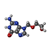  Mass: 225.205 Da / Num. of mol.: 3 / Source method: obtained synthetically / Formula: C8H11N5O3 / Comment: medication, antivirus*YM Mass: 225.205 Da / Num. of mol.: 3 / Source method: obtained synthetically / Formula: C8H11N5O3 / Comment: medication, antivirus*YM#4: Water | ChemComp-HOH / |  Mass: 18.015 Da / Num. of mol.: 388 / Source method: isolated from a natural source / Formula: H2O Mass: 18.015 Da / Num. of mol.: 388 / Source method: isolated from a natural source / Formula: H2O |
|---|
-Experimental details
-Experiment
| Experiment | Method: X-RAY DIFFRACTION / Number of used crystals: 1 |
|---|
- Sample preparation
| Crystal | Density Matthews: 1.79 Å3/Da / Density % sol: 31.14 % |
|---|---|
| Crystal grow | Temperature: 293 K / Method: vapor diffusion, hanging drop / pH: 8 / Details: 100 mM Tris, pH 8.0, 25%PEG 3350, and 25 mM MgCl2 |
-Data collection
| Diffraction | Mean temperature: 100 K |
|---|---|
| Diffraction source | Source: SYNCHROTRON / Site: LNLS  / Beamline: D03B-MX1 / Wavelength: 1.427 Å / Beamline: D03B-MX1 / Wavelength: 1.427 Å |
| Detector | Type: MAR scanner 300 mm plate / Detector: CCD / Date: Feb 2, 2009 |
| Radiation | Monochromator: GRAPHITE / Protocol: SINGLE WAVELENGTH / Monochromatic (M) / Laue (L): M / Scattering type: x-ray |
| Radiation wavelength | Wavelength: 1.427 Å / Relative weight: 1 |
| Reflection | Resolution: 2.1→33.94 Å / Num. all: 39559 / Num. obs: 35559 / % possible obs: 90 % / Observed criterion σ(F): 2 / Observed criterion σ(I): 2 / Redundancy: 7 % / Biso Wilson estimate: 20 Å2 / Rmerge(I) obs: 0.08 / Rsym value: 0.08 / Net I/σ(I): 10 |
| Reflection shell | Resolution: 2.1→2.2 Å / Redundancy: 4 % / Rmerge(I) obs: 0.2 / Mean I/σ(I) obs: 3 / Num. unique all: 2200 / Rsym value: 0.2 / % possible all: 90 |
- Processing
| Software |
| ||||||||||||||||||||||||||||||||||||||||||||||||||||||||||||||||||||||||||||||||||||||||||
|---|---|---|---|---|---|---|---|---|---|---|---|---|---|---|---|---|---|---|---|---|---|---|---|---|---|---|---|---|---|---|---|---|---|---|---|---|---|---|---|---|---|---|---|---|---|---|---|---|---|---|---|---|---|---|---|---|---|---|---|---|---|---|---|---|---|---|---|---|---|---|---|---|---|---|---|---|---|---|---|---|---|---|---|---|---|---|---|---|---|---|---|
| Refinement | Method to determine structure: MOLECULAR REPLACEMENT Starting model: 1N3I Resolution: 2.1→33.94 Å / Cor.coef. Fo:Fc: 0.945 / Cor.coef. Fo:Fc free: 0.876 / Cross valid method: THROUGHOUT / σ(F): 2 / σ(I): 2 / ESU R: 0.369 / ESU R Free: 0.251 / Stereochemistry target values: MAXIMUM LIKELIHOOD / Details: HYDROGENS HAVE BEEN ADDED IN THE RIDING POSITIONS
| ||||||||||||||||||||||||||||||||||||||||||||||||||||||||||||||||||||||||||||||||||||||||||
| Solvent computation | Ion probe radii: 0.8 Å / Shrinkage radii: 0.8 Å / VDW probe radii: 1.2 Å / Solvent model: MASK | ||||||||||||||||||||||||||||||||||||||||||||||||||||||||||||||||||||||||||||||||||||||||||
| Displacement parameters | Biso mean: 23.187 Å2
| ||||||||||||||||||||||||||||||||||||||||||||||||||||||||||||||||||||||||||||||||||||||||||
| Refine analyze | Luzzati coordinate error obs: 0.03 Å | ||||||||||||||||||||||||||||||||||||||||||||||||||||||||||||||||||||||||||||||||||||||||||
| Refinement step | Cycle: LAST / Resolution: 2.1→33.94 Å
| ||||||||||||||||||||||||||||||||||||||||||||||||||||||||||||||||||||||||||||||||||||||||||
| Refine LS restraints |
| ||||||||||||||||||||||||||||||||||||||||||||||||||||||||||||||||||||||||||||||||||||||||||
| LS refinement shell | Resolution: 2.1→2.155 Å / Total num. of bins used: 20
|
