 Movie
Movie Controller

[English] 日本語
 Yorodumi
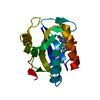
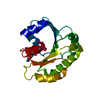
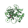
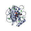
- PDB-3ip0: Crystal structure of E. coli HPPK in complex with MgAMPCPP and 6-... -
+ Open data

- Basic information
| Entry | Database: PDB / ID: 3ip0 | |||||||||
|---|---|---|---|---|---|---|---|---|---|---|
| Title | Crystal structure of E. coli HPPK in complex with MgAMPCPP and 6-hydroxymethylpterin/6-carboxypterin | |||||||||
 Components Components | 2-amino-4-hydroxy-6-hydroxymethyldihydropteridine pyrophosphokinase | |||||||||
 Keywords Keywords | TRANSFERASE / alpha beta / ATP-binding / Folate biosynthesis / Kinase / Nucleotide-binding | |||||||||
| Function / homology |  Function and homology information Function and homology information2-amino-4-hydroxy-6-hydroxymethyldihydropteridine diphosphokinase / 2-amino-4-hydroxy-6-hydroxymethyldihydropteridine diphosphokinase activity / folic acid biosynthetic process / tetrahydrofolate biosynthetic process / kinase activity / magnesium ion binding / ATP binding Similarity search - Function | |||||||||
| Biological species |  | |||||||||
| Method |  X-RAY DIFFRACTION / SYNCHROTRON / FOURIER SYNTHESIS / Resolution: 0.89 Å X-RAY DIFFRACTION / SYNCHROTRON / FOURIER SYNTHESIS / Resolution: 0.89 Å | |||||||||
 Authors Authors | Blaszczyk, J. / Ji, X. | |||||||||
 Citation Citation | Journal: Biochemistry / Year: 2003 Title: Dynamic roles of arginine residues 82 and 92 of Escherichia coli 6-hydroxymethyl-7,8-dihydroptein pyrophosphokinase: Crystallographic studies Authors: Blaszczyk, J. / Li, Y. / Shi, G. / Yan, H. / Ji, X. #1: Journal: Structure / Year: 2000Title: Catalytic center assembly of HPPK as revealed by the crystal structure of a ternary complex at 1.25-angstrom resolution Authors: Blaszczyk, J. / Shi, G. / Yan, H. / Ji, X. | |||||||||
| History |
|
- Structure visualization
| Structure viewer | Molecule: MolmilJmol/JSmol |
|---|
- Downloads & links
-Download
| PDBx/mmCIF format | 3ip0.cif.gz | 127.2 KB | Display | PDBx/mmCIF format |
|---|---|---|---|---|
| PDB format | pdb3ip0.ent.gz | 99 KB | Display | PDB format |
| PDBx/mmJSON format | 3ip0.json.gz | Tree view | PDBx/mmJSON format | |
| Others |  Other downloads Other downloads |
-Validation report
| Arichive directory | https://data.pdbj.org/pub/pdb/validation_reports/ip/3ip0ftp://data.pdbj.org/pub/pdb/validation_reports/ip/3ip0 | HTTPS FTP |
|---|
-Related structure data
| Related structure data |  1f9hC  1g4cC  1hq2C  1im6C  1kbrC  1q0nS S: Starting model for refinement C: citing same article ( |
|---|---|
| Similar structure data |


-Links
 PDBj
PDBj


- Assembly
| Deposited unit | 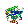
| ||||||||||||||||||
|---|---|---|---|---|---|---|---|---|---|---|---|---|---|---|---|---|---|---|---|
| 1 |
| ||||||||||||||||||
| 2 | 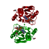
| ||||||||||||||||||
| Unit cell |
| ||||||||||||||||||
| Components on special symmetry positions |
|
-Components
-Protein , 1 types, 1 molecules A
| #1: Protein | Mass: 17966.535 Da / Num. of mol.: 1 Source method: isolated from a genetically manipulated source Source: (gene. exp.) References: UniProt: P26281, 2-amino-4-hydroxy-6-hydroxymethyldihydropteridine diphosphokinase |
|---|
-Non-polymers , 7 types, 337 molecules 

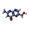
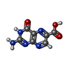


| #2: Chemical |  Mass: 24.305 Da / Num. of mol.: 2 / Source method: obtained synthetically / Formula: Mg Mass: 24.305 Da / Num. of mol.: 2 / Source method: obtained synthetically / Formula: Mg#3: Chemical | ChemComp-APC / |  Mass: 505.208 Da / Num. of mol.: 1 / Source method: obtained synthetically / Formula: C11H18N5O12P3 / Comment: AMP-CPP, energy-carrying molecule analogue*YM Mass: 505.208 Da / Num. of mol.: 1 / Source method: obtained synthetically / Formula: C11H18N5O12P3 / Comment: AMP-CPP, energy-carrying molecule analogue*YM#4: Chemical | ChemComp-HHR / |  Mass: 193.163 Da / Num. of mol.: 1 / Source method: obtained synthetically / Formula: C7H7N5O2 Mass: 193.163 Da / Num. of mol.: 1 / Source method: obtained synthetically / Formula: C7H7N5O2#5: Chemical | ChemComp-HHS / |  Mass: 207.146 Da / Num. of mol.: 1 / Source method: obtained synthetically / Formula: C7H5N5O3 Mass: 207.146 Da / Num. of mol.: 1 / Source method: obtained synthetically / Formula: C7H5N5O3#6: Chemical |  Mass: 35.453 Da / Num. of mol.: 3 / Source method: obtained synthetically / Formula: Cl Mass: 35.453 Da / Num. of mol.: 3 / Source method: obtained synthetically / Formula: Cl#7: Chemical | ChemComp-ACT /  Mass: 59.044 Da / Num. of mol.: 4 / Source method: obtained synthetically / Formula: C2H3O2 Mass: 59.044 Da / Num. of mol.: 4 / Source method: obtained synthetically / Formula: C2H3O2#8: Water | ChemComp-HOH / | Mass: 18.015 Da / Num. of mol.: 325 / Source method: isolated from a natural source / Formula: H2O |
|---|
-Experimental details
-Experiment
| Experiment | Method: X-RAY DIFFRACTION / Number of used crystals: 1 |
|---|
- Sample preparation
| Crystal | Density Matthews: 1.99 Å3/Da / Density % sol: 38.33 % |
|---|---|
| Crystal grow | Temperature: 292 K / Method: vapor diffusion, hanging drop / pH: 4.6 Details: PEG 4000, Ammonium acetate, Sodium acetate, Glycerol., pH 4.6, VAPOR DIFFUSION, HANGING DROP, temperature 292K |
-Data collection
| Diffraction | Mean temperature: 100 K |
|---|---|
| Diffraction source | Source: SYNCHROTRON / Site: NSLS  / Beamline: X9B / Wavelength: 1.00928 Å / Beamline: X9B / Wavelength: 1.00928 Å |
| Detector | Type: ADSC QUANTUM 4 / Detector: CCD / Date: Oct 18, 1999 / Details: mirrors |
| Radiation | Monochromator: Silicon 111 / Protocol: SINGLE WAVELENGTH / Monochromatic (M) / Laue (L): M / Scattering type: x-ray |
| Radiation wavelength | Wavelength: 1.00928 Å / Relative weight: 1 |
| Reflection | Resolution: 0.89→40 Å / Num. all: 100866 / Num. obs: 100866 / % possible obs: 93.1 % / Observed criterion σ(F): -6 / Observed criterion σ(I): -3 / Redundancy: 4.44 % / Biso Wilson estimate: 6.8 Å2 / Rmerge(I) obs: 0.043 / Χ2: 1.003 / Net I/σ(I): 14.5 |
| Reflection shell | Resolution: 0.89→0.92 Å / Redundancy: 2.74 % / Rmerge(I) obs: 0.581 / Mean I/σ(I) obs: 1.73 / Num. unique all: 8139 / Χ2: 1.001 / % possible all: 75.6 |
- Processing
| Software |
| ||||||||||||||||||||||||||||||||||||||||||||||||||||||||||||||||||||||||||||||||||||||||
|---|---|---|---|---|---|---|---|---|---|---|---|---|---|---|---|---|---|---|---|---|---|---|---|---|---|---|---|---|---|---|---|---|---|---|---|---|---|---|---|---|---|---|---|---|---|---|---|---|---|---|---|---|---|---|---|---|---|---|---|---|---|---|---|---|---|---|---|---|---|---|---|---|---|---|---|---|---|---|---|---|---|---|---|---|---|---|---|---|---|
| Refinement | Method to determine structure: FOURIER SYNTHESIS Starting model: PDB entry 1Q0N Resolution: 0.89→23.202 Å / Occupancy max: 1 / Occupancy min: 0.17 / SU ML: 0.06 Isotropic thermal model: Anisotropic for fully-occupied non-hydrogen atoms Cross valid method: Throughout before the final 7 cycles of refinement σ(F): 1.35 / Stereochemistry target values: ML Details: The structure was refined for a total of 49 cycles, including 6 cycles with CNS, 20 cycles with SHELXL and 23 cycles with PHENIX
| ||||||||||||||||||||||||||||||||||||||||||||||||||||||||||||||||||||||||||||||||||||||||
| Solvent computation | Shrinkage radii: 0.9 Å / VDW probe radii: 1.11 Å / Solvent model: FLAT BULK SOLVENT MODEL / Bsol: 75.633 Å2 / ksol: 0.447 e/Å3 | ||||||||||||||||||||||||||||||||||||||||||||||||||||||||||||||||||||||||||||||||||||||||
| Displacement parameters | Biso max: 91.76 Å2 / Biso mean: 12.849 Å2 / Biso min: 3.12 Å2
| ||||||||||||||||||||||||||||||||||||||||||||||||||||||||||||||||||||||||||||||||||||||||
| Refine analyze | Luzzati coordinate error obs: 0.006 Å | ||||||||||||||||||||||||||||||||||||||||||||||||||||||||||||||||||||||||||||||||||||||||
| Refinement step | Cycle: LAST / Resolution: 0.89→23.202 Å
| ||||||||||||||||||||||||||||||||||||||||||||||||||||||||||||||||||||||||||||||||||||||||
| Refine LS restraints |
| ||||||||||||||||||||||||||||||||||||||||||||||||||||||||||||||||||||||||||||||||||||||||
| LS refinement shell | Refine-ID: X-RAY DIFFRACTION / Total num. of bins used: 10
|
