[English] 日本語
Yorodumi
- PDB-3ioi: Crystal structure of the Fucosylgalactoside alpha N-acetylgalacto... -

+
Open data


ID or keywords:

Loading...

-
Basic information

Entry
Database: PDB / ID: 3ioi
Title
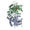
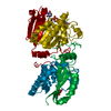
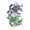
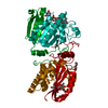
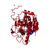
Crystal structure of the Fucosylgalactoside alpha N-acetylgalactosaminyltransferase (GTA, cisAB mutant L266G, G268A) in complex with a novel UDP-Gal derived inhibitor (1GW)
ComponentsHisto-blood group ABO system transferase
KeywordsTRANSFERASE / GTA / ABO / cisAB mutant / AA(Gly)B / ROSSMANN FOLD / INHIBITOR / SEMI-CLOSED CONFORMATION / BLOOD GROUP ANTIGEN / GLYCOPROTEIN / GLYCOSYLTRANSFERASE / GOLGI APPARATUS / MANGANESE / MEMBRANE / METAL-BINDING / POLYMORPHISM / SECRETED / SIGNAL-ANCHOR / TRANSMEMBRANE
Function / homology
Function and homology information


fucosylgalactoside 3-alpha-galactosyltransferase / glycoprotein-fucosylgalactoside alpha-N-acetylgalactosaminyltransferase / glycoprotein-fucosylgalactoside alpha-N-acetylgalactosaminyltransferase activity / fucosylgalactoside 3-alpha-galactosyltransferase activity / ABO blood group biosynthesis / lipid glycosylation / Golgi cisterna membrane / protein glycosylation / antigen binding / manganese ion binding ...fucosylgalactoside 3-alpha-galactosyltransferase / glycoprotein-fucosylgalactoside alpha-N-acetylgalactosaminyltransferase / glycoprotein-fucosylgalactoside alpha-N-acetylgalactosaminyltransferase activity / fucosylgalactoside 3-alpha-galactosyltransferase activity / ABO blood group biosynthesis / lipid glycosylation / Golgi cisterna membrane / protein glycosylation / antigen binding / manganese ion binding / vesicle / carbohydrate metabolic process / Golgi membrane / nucleotide binding / Golgi apparatus / extracellular region
Similarity search - Function
Glycosyl transferase, family 6 / Glycosyltransferase family 6 / Spore Coat Polysaccharide Biosynthesis Protein SpsA; Chain A / Spore Coat Polysaccharide Biosynthesis Protein SpsA; Chain A / Nucleotide-diphospho-sugar transferases / Alpha-Beta Complex / Alpha Beta
Similarity search - Domain/homology
Chem-1GW / : / Histo-blood group ABO system transferase
Similarity search - Component
Biological speciesHomo sapiens (human)
MethodX-RAY DIFFRACTION / SYNCHROTRON / MOLECULAR REPLACEMENT / molecular replacement / Resolution: 1.45 Å
AuthorsPesnot, T. / Jorgensen, R. / Palcic, M.M. / Wagner, G.K.
CitationJournal: Nat.Chem.Biol. / Year: 2010
Title: Structural and mechanistic basis for a new mode of glycosyltransferase inhibition.
Authors: Pesnot, T. / Jorgensen, R. / Palcic, M.M. / Wagner, G.K.
History
DepositionAug 14, 2009Deposition site: RCSB / Processing site: RCSB
Revision 1.0Apr 7, 2010Provider: repository / Type: Initial release
Revision 1.1Jul 13, 2011Group: Version format compliance
Revision 1.2Nov 12, 2014Group: Structure summary
Revision 1.3Sep 27, 2017Group: Data collection / Category: diffrn_detector / diffrn_source
Item: _diffrn_detector.detector / _diffrn_source.pdbx_synchrotron_site
Revision 1.4Oct 13, 2021Group: Database references / Derived calculations
Category: database_2 / pdbx_struct_conn_angle ...database_2 / pdbx_struct_conn_angle / struct_conn / struct_ref_seq_dif / struct_site
Item: _database_2.pdbx_DOI / _database_2.pdbx_database_accession ..._database_2.pdbx_DOI / _database_2.pdbx_database_accession / _pdbx_struct_conn_angle.ptnr1_auth_comp_id / _pdbx_struct_conn_angle.ptnr1_auth_seq_id / _pdbx_struct_conn_angle.ptnr1_label_asym_id / _pdbx_struct_conn_angle.ptnr1_label_atom_id / _pdbx_struct_conn_angle.ptnr1_label_comp_id / _pdbx_struct_conn_angle.ptnr1_label_seq_id / _pdbx_struct_conn_angle.ptnr3_auth_comp_id / _pdbx_struct_conn_angle.ptnr3_auth_seq_id / _pdbx_struct_conn_angle.ptnr3_label_asym_id / _pdbx_struct_conn_angle.ptnr3_label_atom_id / _pdbx_struct_conn_angle.ptnr3_label_comp_id / _pdbx_struct_conn_angle.ptnr3_label_seq_id / _pdbx_struct_conn_angle.value / _struct_conn.pdbx_dist_value / _struct_conn.ptnr1_auth_comp_id / _struct_conn.ptnr1_auth_seq_id / _struct_conn.ptnr1_label_asym_id / _struct_conn.ptnr1_label_atom_id / _struct_conn.ptnr1_label_comp_id / _struct_conn.ptnr1_label_seq_id / _struct_conn.ptnr2_auth_comp_id / _struct_conn.ptnr2_auth_seq_id / _struct_conn.ptnr2_label_asym_id / _struct_conn.ptnr2_label_atom_id / _struct_conn.ptnr2_label_comp_id / _struct_conn.ptnr2_label_seq_id / _struct_ref_seq_dif.details / _struct_site.pdbx_auth_asym_id / _struct_site.pdbx_auth_comp_id / _struct_site.pdbx_auth_seq_id
Revision 1.5Sep 6, 2023Group: Data collection / Refinement description
Category: chem_comp_atom / chem_comp_bond / pdbx_initial_refinement_model

-
Structure visualization

Structure viewerMolecule:
MolmilJmol/JSmol

Downloads & links

-
Assembly

Deposited unit
A: Histo-blood group ABO system transferase
hetero molecules


Theoretical massNumber of molelcules
Total (without water)35,4814
Polymers34,6541
Non-polymers8273
Water4,035224
1
A: Histo-blood group ABO system transferase
hetero molecules

A: Histo-blood group ABO system transferase
hetero molecules


Theoretical massNumber of molelcules
Total (without water)70,9638
Polymers69,3082
Non-polymers1,6556
Water362
TypeNameSymmetry operationNumber
identity operation1_555x,y,z1
crystal symmetry operation3_554-x,y,-z-1/21
Buried area5280 Å2
ΔGint-59 kcal/mol
Surface area22660 Å2
MethodPISA
Unit cell
Length a, b, c (Å)52.160, 148.440, 79.000
Angle α, β, γ (deg.)90.000, 90.000, 90.000
Int Tables number20
Space group name H-MC2221

-
Components

#1: Protein Histo-blood group ABO system transferase / NAGAT / Glycoprotein-fucosylgalactoside alpha-N-acetylgalactosaminyltransferase / ...NAGAT / Glycoprotein-fucosylgalactoside alpha-N-acetylgalactosaminyltransferase / Fucosylglycoprotein alpha-N-acetylgalactosaminyltransferase / Histo-blood group A transferase / A transferase / Glycoprotein-fucosylgalactoside alpha-galactosyltransferase / Fucosylglycoprotein 3-alpha-galactosyltransferase / Histo-blood group B transferase / B transferase / Fucosylglycoprotein alpha-N-acetylgalactosaminyltransferase soluble form


Mass: 34654.008 Da / Num. of mol.: 1 / Fragment: Extracellular catalytic domain / Mutation: L266G, G268A
Source method: isolated from a genetically manipulated source
Source: (gene. exp.) Homo sapiens (human) / Gene: ABO / Plasmid: PCW DELTA 1AC / Production host: Escherichia coli BL21 (bacteria) / Strain (production host): BL21
References: UniProt: P16442, glycoprotein-fucosylgalactoside alpha-N-acetylgalactosaminyltransferase, fucosylgalactoside 3-alpha-galactosyltransferase
#2: Chemical ChemComp-MN / MANGANESE (II) ION


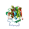
Mass: 54.938 Da / Num. of mol.: 1 / Source method: obtained synthetically / Formula: Mn
#3: Chemical ChemComp-SO4 / SULFATE ION / Sulfate


Mass: 96.063 Da / Num. of mol.: 1 / Source method: obtained synthetically / Formula: SO4
#4: Chemical ChemComp-1GW / 5-(2-FORMYLTHIEN-5-YL)-URIDINE-5'-DIPHOSPHATE-ALPHA-D-GALACTOSE


Mass: 676.436 Da / Num. of mol.: 1 / Source method: obtained synthetically / Formula: C20H26N2O18P2S
#5: Water ChemComp-HOH / water / Water


Mass: 18.015 Da / Num. of mol.: 224 / Source method: isolated from a natural source / Formula: H2O

-
Experimental details

-
Experiment

ExperimentMethod: X-RAY DIFFRACTION / Number of used crystals: 1

-
Sample preparation

CrystalDensity Matthews: 2.21 Å3/Da / Density % sol: 44.25 %
Crystal growTemperature: 292 K / Method: vapor diffusion, hanging drop / pH: 7
Details: 50 mM MOPS pH 7, 50-200 mM ammonium sulfate, 50 mM MnCl2, and 6-9% PEG-3350, VAPOR DIFFUSION, HANGING DROP, temperature 292K

-
Data collection

DiffractionMean temperature: 100 K
Diffraction sourceSource: SYNCHROTRON / Site: MAX II / Beamline: I911-5 / Wavelength: 0.90772 Å
DetectorType: MAR555 FLAT PANEL / Detector: IMAGE PLATE / Date: May 29, 2009
Details: Multilayer mirror, curved to focus in the vertical (R = 400 m)
RadiationMonochromator: Bent Si (220) crystal, horizontally focusing / Protocol: SINGLE WAVELENGTH / Monochromatic (M) / Laue (L): M / Scattering type: x-ray
Radiation wavelengthWavelength: 0.90772 Å / Relative weight: 1
ReflectionResolution: 1.45→20 Å / Num. obs: 54487 / % possible obs: 99.6 % / Observed criterion σ(I): -3 / Redundancy: 11.07 % / Biso Wilson estimate: 22.001 Å2 / Rmerge(I) obs: 0.084 / Net I/σ(I): 17.34
Reflection shellResolution: 1.45→1.6 Å / Redundancy: 7.25 % / Rmerge(I) obs: 0.681 / Mean I/σ(I) obs: 2.9 / Num. measured obs: 99444 / Num. unique all: 13715 / Num. unique obs: 13715 / % possible all: 99.3

-
Phasing

PhasingMethod: molecular replacement
Phasing MRModel details: Phaser MODE: MR_AUTO
Highest resolutionLowest resolution
Rotation2.5 Å19.75 Å
Translation2.5 Å19.75 Å

-
Processing

Software
NameVersionClassificationNB
XSCALEdata scaling
PHASER1.3.3phasing
PHENIXrefinement
PDB_EXTRACT3.005data extraction
MAR345dtbdata collection
XDSdata reduction
RefinementMethod to determine structure: MOLECULAR REPLACEMENT
Starting model: 2RIT
Resolution: 1.45→19.644 Å / Occupancy max: 1 / Occupancy min: 0.35 / FOM work R set: 0.907 / SU ML: 0.03 / Isotropic thermal model: Isotropic / Cross valid method: THROUGHOUT / σ(F): 1.99 / Stereochemistry target values: ML
RfactorNum. reflection% reflectionSelection details
Rfree0.175 1634 3 %RANDOM
Rwork0.159 ---
obs0.16 54482 99.61 %-
Solvent computationShrinkage radii: 0.9 Å / VDW probe radii: 1.11 Å / Solvent model: FLAT BULK SOLVENT MODEL / Bsol: 69.529 Å2 / ksol: 0.46 e/Å3
Displacement parametersBiso max: 109.67 Å2 / Biso mean: 20.78 Å2 / Biso min: 6.4 Å2
Baniso -1Baniso -2Baniso -3
1--1.616 Å20 Å20 Å2
2--0.012 Å2-0 Å2
3---1.605 Å2
Refinement stepCycle: LAST / Resolution: 1.45→19.644 Å
ProteinNucleic acidLigandSolventTotal
Num. atoms2241 0 49 224 2514
Refine LS restraints
Refine-IDTypeDev idealNumber
X-RAY DIFFRACTIONf_bond_d0.012408
X-RAY DIFFRACTIONf_angle_d1.4183286
X-RAY DIFFRACTIONf_chiral_restr0.084349
X-RAY DIFFRACTIONf_plane_restr0.007413
X-RAY DIFFRACTIONf_dihedral_angle_d17.619931
LS refinement shell

Refine-ID: X-RAY DIFFRACTION / Total num. of bins used: 12

Resolution (Å)Rfactor RfreeNum. reflection RfreeRfactor RworkNum. reflection RworkNum. reflection all% reflection obs (%)
1.45-1.4930.271340.2454292442699
1.493-1.5410.2651330.224360449399
1.541-1.5960.2281350.18243534488100
1.596-1.660.1981330.16743664499100
1.66-1.7350.1691370.15643974534100
1.735-1.8270.1841370.14743894526100
1.827-1.9410.1731350.14243844519100
1.941-2.0910.1541360.13144164552100
2.091-2.3010.1341370.13244144551100
2.301-2.6330.1611380.14444564594100
2.633-3.3150.1781380.15644714609100
3.315-19.6460.1621410.1664550469198
Refinement TLS params.

Method: refined / Refine-ID: X-RAY DIFFRACTION

IDL112)L122)L132)L222)L232)L332)S11 (Å °)S12 (Å °)S13 (Å °)S21 (Å °)S22 (Å °)S23 (Å °)S31 (Å °)S32 (Å °)S33 (Å °)T112)T122)T132)T222)T232)T332)Origin x (Å)Origin y (Å)Origin z (Å)
10.1092-0.5650.45541.6673-0.29071.29120.2662-0.049-0.03480.0549-0.2679-0.30180.13350.3501-0.00520.16830.02880.00720.2191-0.00150.18898.643511.2313-31.8055
20.79270.2755-0.1386-0.27440.1290.7632-0.02250.0271-0.12720.00890.0396-0.08230.1578-0.0405-0.01220.0955-0.0018-0.00980.0649-0.00510.10861.67939.3421-15.4419
31.75770.82190.27230.94890.0970.0641-0.06060.2613-0.0641-0.06380.11910.002-0.02180.0144-0.05650.08420.0037-0.00050.12440.01390.09313.465721.3069-24.2778
40.99370.07860.1670.552-0.27521.16250.0743-0.15890.01680.070.0286-0.087-0.0309-0.0114-0.06040.0955-0.0237-0.01840.1149-0.00630.084120.762224.40260.3758
50.14830.22980.03970.65440.07681.48590.04850.0932-0.0774-0.0139-0.0103-0.1677-0.03830.171-0.02810.082-0.0002-0.00520.10390.00110.116123.279720.5642-11.1499
60.3841-0.2003-0.09521.04250.3125.220.07380.07730.0203-0.15090.0977-0.1772-0.40360.1965-0.17830.1073-0.01590.02270.0907-0.02380.09820.460530.3913-9.1818
73.9462.1690.1781.90530.07610.54130.1757-0.1479-0.03960.3123-0.0891-0.2116-0.33710.2468-0.07960.1932-0.0449-0.00850.1625-0.01870.136326.663929.96283.8832
81.0403-0.6094-0.70990.85530.00021.38040.1178-0.23930.17460.0937-0.1141-0.0131-0.09380.4187-0.02020.0788-0.01560.0040.1749-0.01050.079227.704928.0184-12.0958
91.49970.1399-0.93890.8692-0.38830.0439-0.0014-0.19870.06840.28490.18630.3049-0.1529-0.0206-0.14440.26340.01040.0480.19530.00020.180216.791532.11335.1799
102.0904-1.01220.55111.60181.05041.16320.4056-0.62730.744-0.1065-0.2007-0.1273-0.1777-0.0567-0.12860.239-0.01330.0670.3615-0.15170.38779.203234.94481.8048
113.4052-0.3623-0.92684.35634.68662.08640.0789-0.50080.8066-0.3213-0.0349-0.0884-0.4781-0.1234-0.02050.15950.00620.00870.1464-0.04790.242311.925436.1419-9.525
120.3810.21850.2804-0.0124-0.17990.91640.06770.0024-0.07720.0057-0.0484-0.00490.12880.0774-0.02530.1160.0004-0.02420.09420.02130.116118.3816.2408-4.837
130.76830.0108-0.25320.14690.22880.150.0185-0.0458-0.16280.086-0.0535-0.01640.0432-0.02360.03820.0955-0.0059-0.01110.07860.01380.10484.435613.8522-6.7239
140.96640.2376-0.45121.75220.77791.28270.0751-0.3326-0.00810.3454-0.11220.37870.13-0.39040.01750.1674-0.03120.04770.22070.02290.1479-7.190116.38346.8792
150.61480.1444-0.04630.29190.25780.09860.0492-0.0707-0.01310.0507-0.045-0.01020.0506-0.0318-0.0070.1009-0.0116-0.0060.0990.01620.0943.906919.0479-4.5602
161.1444-0.6734-0.95840.72790.12961.17390.15850.11840.24820.0264-0.0869-0.0324-0.37-0.2789-0.07710.15030.02420.02020.1319-0.00450.13081.637232.0702-2.6281
170.22520.40930.47062.11030.0826-3.51690.0466-0.29970.07030.88040.1529-0.1310.01170.1249-0.1620.3397-00.03120.2468-0.04520.1097-1.409128.619812.7101
181.0109-0.085-0.23190.8064-0.21140.0490.0354-0.1774-0.06790.14530.0070.0137-0.0335-0.0995-0.02910.0967-0.00260.00130.13490.01740.0831-0.48920.4469-1.2064
190.2456-0.37450.1475-0.246-0.10291.3099-0.02690.026-0.12380.0767-0.07340.02490.2150.01110.06830.1561-0.0063-0.02090.07960.03110.133413.4695.9194-4.5662
201.13090.06330.8358-0.0982-0.49773.13060.0781-0.0469-0.25220.0514-0.1373-0.14230.26640.16270.00170.1240.0053-0.04160.10830.02340.138317.08839.5764-5.4206
Refinement TLS group
IDRefine-IDRefine TLS-IDSelection detailsAuth asym-IDAuth seq-ID
1X-RAY DIFFRACTION1(chain A and resid 65:80)A65 - 80
2X-RAY DIFFRACTION2(chain A and resid 81:97)A81 - 97
3X-RAY DIFFRACTION3(chain A and resid 98:113)A98 - 113
4X-RAY DIFFRACTION4(chain A and resid 114:132)A114 - 132
5X-RAY DIFFRACTION5(chain A and resid 133:145)A133 - 145
6X-RAY DIFFRACTION6(chain A and resid 146:151)A146 - 151
7X-RAY DIFFRACTION7(chain A and resid 152:161)A152 - 161
8X-RAY DIFFRACTION8(chain A and resid 162:169)A162 - 169
9X-RAY DIFFRACTION9(chain A and resid 170:188)A170 - 188
10X-RAY DIFFRACTION10(chain A and resid 189:196)A189 - 196
11X-RAY DIFFRACTION11(chain A and resid 197:207)A197 - 207
12X-RAY DIFFRACTION12(chain A and resid 208:219)A208 - 219
13X-RAY DIFFRACTION13(chain A and resid 220:242)A220 - 242
14X-RAY DIFFRACTION14(chain A and resid 243:260)A243 - 260
15X-RAY DIFFRACTION15(chain A and resid 261:274)A261 - 274
16X-RAY DIFFRACTION16(chain A and resid 275:286)A275 - 286
17X-RAY DIFFRACTION17(chain A and resid 287:298)A287 - 298
18X-RAY DIFFRACTION18(chain A and resid 299:319)A299 - 319
19X-RAY DIFFRACTION19(chain A and resid 320:335)A320 - 335
20X-RAY DIFFRACTION20(chain A and resid 336:346)A336 - 346

+
About Yorodumi

-
News

-
Feb 9, 2022. New format data for meta-information of EMDB entries

New format data for meta-information of EMDB entries

  • Version 3 of the EMDB header file is now the official format.
  • The previous official version 1.9 will be removed from the archive.

Related info.:EMDB header

External links:wwPDB to switch to version 3 of the EMDB data model

-
Aug 12, 2020. Covid-19 info

Covid-19 info

URL: https://pdbj.org/emnavi/covid19.php

New page: Covid-19 featured information page in EM Navigator.

Related info.:Covid-19 info / Mar 5, 2020. Novel coronavirus structure data

+
Mar 5, 2020. Novel coronavirus structure data

Novel coronavirus structure data

Related info.:Yorodumi Speices / Aug 12, 2020. Covid-19 info

External links:COVID-19 featured content - PDBj / Molecule of the Month (242):Coronavirus Proteases

+
Jan 31, 2019. EMDB accession codes are about to change! (news from PDBe EMDB page)

EMDB accession codes are about to change! (news from PDBe EMDB page)

  • The allocation of 4 digits for EMDB accession codes will soon come to an end. Whilst these codes will remain in use, new EMDB accession codes will include an additional digit and will expand incrementally as the available range of codes is exhausted. The current 4-digit format prefixed with “EMD-” (i.e. EMD-XXXX) will advance to a 5-digit format (i.e. EMD-XXXXX), and so on. It is currently estimated that the 4-digit codes will be depleted around Spring 2019, at which point the 5-digit format will come into force.
  • The EM Navigator/Yorodumi systems omit the EMD- prefix.

Related info.:Q: What is EMD? / ID/Accession-code notation in Yorodumi/EM Navigator

External links:EMDB Accession Codes are Changing Soon! / Contact to PDBj

+
Jul 12, 2017. Major update of PDB

Major update of PDB

  • wwPDB released updated PDB data conforming to the new PDBx/mmCIF dictionary.
  • This is a major update changing the version number from 4 to 5, and with Remediation, in which all the entries are updated.
  • In this update, many items about electron microscopy experimental information are reorganized (e.g. em_software).
  • Now, EM Navigator and Yorodumi are based on the updated data.

External links:wwPDB Remediation / Enriched Model Files Conforming to OneDep Data Standards Now Available in the PDB FTP Archive

-
Yorodumi

Thousand views of thousand structures

  • Yorodumi is a browser for structure data from EMDB, PDB, SASBDB, etc.
  • This page is also the successor to EM Navigator detail page, and also detail information page/front-end page for Omokage search.
  • The word "yorodu" (or yorozu) is an old Japanese word meaning "ten thousand". "mi" (miru) is to see.

Related info.:EMDB / PDB / SASBDB / Comparison of 3 databanks / Yorodumi Search / Aug 31, 2016. New EM Navigator & Yorodumi / Yorodumi Papers / Jmol/JSmol / Function and homology information / Changes in new EM Navigator and Yorodumi

Read more