| 登録情報 | データベース: PDB / ID: 3im3
|
|---|
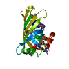
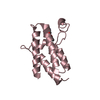
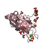
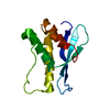
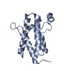
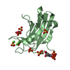
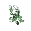
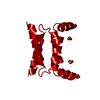
| タイトル | Crystal structure of PKA RI alpha dimerization/docking domain |
|---|
 要素 要素 | cAMP-dependent protein kinase type I-alpha regulatory subunit |
|---|
 キーワード キーワード | STRUCTURAL PROTEIN / SIGNALING PROTEIN / helix-turn-helix / Acetylation / cAMP / cAMP-binding / Disulfide bond / Nucleotide-binding / Phosphoprotein |
|---|
| 機能・相同性 |  機能・相同性情報 機能・相同性情報
sperm head-tail coupling apparatus / PKA activation in glucagon signalling / DARPP-32 events / CREB1 phosphorylation through the activation of Adenylate Cyclase / GPER1 signaling / Factors involved in megakaryocyte development and platelet production / PKA activation / nucleotide-activated protein kinase complex / Hedgehog 'off' state / negative regulation of cAMP/PKA signal transduction ...sperm head-tail coupling apparatus / PKA activation in glucagon signalling / DARPP-32 events / CREB1 phosphorylation through the activation of Adenylate Cyclase / GPER1 signaling / Factors involved in megakaryocyte development and platelet production / PKA activation / nucleotide-activated protein kinase complex / Hedgehog 'off' state / negative regulation of cAMP/PKA signal transduction / cAMP-dependent protein kinase inhibitor activity / sarcomere organization / cAMP-dependent protein kinase complex / High laminar flow shear stress activates signaling by PIEZO1 and PECAM1:CDH5:KDR in endothelial cells / Vasopressin regulates renal water homeostasis via Aquaporins / negative regulation of activated T cell proliferation / protein kinase A catalytic subunit binding / mesoderm formation / immunological synapse / plasma membrane raft / axoneme / cardiac muscle cell proliferation / cAMP binding / multivesicular body / cellular response to glucagon stimulus / neuromuscular junction / positive regulation of insulin secretion / adenylate cyclase-activating G protein-coupled receptor signaling pathway / protein domain specific binding / negative regulation of gene expression / ubiquitin protein ligase binding / centrosome / glutamatergic synapse / identical protein binding / cytoplasm / cytosol類似検索 - 分子機能 cAMP-dependent protein kinase regulatory subunit, dimerization-anchoring domain / cAMP-dependent Protein Kinase, Chain A / cAMP-dependent protein kinase regulatory subunit / cAMP-dependent protein kinase regulatory subunit, dimerization-anchoring domain / Regulatory subunit of type II PKA R-subunit / RIIalpha, Regulatory subunit portion of type II PKA R-subunit / : / Cyclic nucleotide-binding domain signature 2. / Cyclic nucleotide-binding domain signature 1. / Cyclic nucleotide-binding, conserved site ...cAMP-dependent protein kinase regulatory subunit, dimerization-anchoring domain / cAMP-dependent Protein Kinase, Chain A / cAMP-dependent protein kinase regulatory subunit / cAMP-dependent protein kinase regulatory subunit, dimerization-anchoring domain / Regulatory subunit of type II PKA R-subunit / RIIalpha, Regulatory subunit portion of type II PKA R-subunit / : / Cyclic nucleotide-binding domain signature 2. / Cyclic nucleotide-binding domain signature 1. / Cyclic nucleotide-binding, conserved site / Cyclic nucleotide-monophosphate binding domain / Cyclic nucleotide-binding domain / cAMP/cGMP binding motif profile. / Cyclic nucleotide-binding domain / Cyclic nucleotide-binding domain superfamily / RmlC-like jelly roll fold / Up-down Bundle / Mainly Alpha類似検索 - ドメイン・相同性 FORMIC ACID / cAMP-dependent protein kinase type I-alpha regulatory subunit類似検索 - 構成要素 |
|---|
| 生物種 |   Bos taurus (ウシ) Bos taurus (ウシ) |
|---|
| 手法 |  X線回折 / シンクロトロン / 単波長異常分散 / 解像度: 2 Å X線回折 / シンクロトロン / 単波長異常分散 / 解像度: 2 Å |
|---|
 データ登録者 データ登録者 | Sarma, G.N. / Kinderman, F.S. / Kim, C. / von Daake, S. / Taylor, S.S. |
|---|
 引用 引用 | ジャーナル: Structure / 年: 2010
タイトル: Structure of D-AKAP2:PKA RI Complex: Insights into AKAP Specificity and Selectivity
著者: Sarma, G.N. / Kinderman, F.S. / Kim, C. / von Daake, S. / Chen, L. / Wang, B.C. / Taylor, S.S. |
|---|
| 履歴 | | 登録 | 2009年8月9日 | 登録サイト: RCSB / 処理サイト: RCSB |
|---|
| 改定 1.0 | 2010年2月2日 | Provider: repository / タイプ: Initial release |
|---|
| 改定 1.1 | 2011年7月13日 | Group: Version format compliance |
|---|
| 改定 1.2 | 2021年4月7日 | Group: Derived calculations / カテゴリ: struct_site
Item: _struct_site.pdbx_auth_asym_id / _struct_site.pdbx_auth_comp_id / _struct_site.pdbx_auth_seq_id |
|---|
| 改定 1.3 | 2024年11月20日 | Group: Data collection / Database references / Structure summary
カテゴリ: chem_comp_atom / chem_comp_bond ...chem_comp_atom / chem_comp_bond / database_2 / pdbx_entry_details / pdbx_modification_feature
Item: _database_2.pdbx_DOI / _database_2.pdbx_database_accession |
|---|
|
|---|
 ムービー
ムービー コントローラー


 データを開く
 基本情報
基本情報 構造の表示
構造の表示 ダウンロードとリンク
 その他のダウンロード

 PDBj
PDBj


 集合体
集合体


 分子量: 46.025 Da / 分子数: 2 / 由来タイプ: 合成 / 式: CH2O2
 分子量: 18.015 Da / 分子数: 29 / 由来タイプ: 天然 / 式: H2O
 試料調製
試料調製 / ビームライン: 22-BM / 波長: 1 Å
 解析
解析