 Movie
Movie Controller

[English] 日本語
 Yorodumi
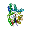
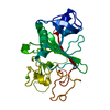
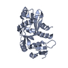
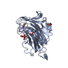
- PDB-3im0: Crystal structure of Chlorella virus vAL-1 soaked in 200mM D-gluc... -
+ Open data

- Basic information
| Entry | Database: PDB / ID: 3im0 | ||||||
|---|---|---|---|---|---|---|---|
| Title | Crystal structure of Chlorella virus vAL-1 soaked in 200mM D-glucuronic acid, 10% PEG-3350, and 200mM glycine-NaOH (pH 10.0) | ||||||
 Components Components | VAL-1 | ||||||
 Keywords Keywords | LYASE / alginate lyase / polysaccharide lyase family 14 / chlorella virus | ||||||
| Function / homology | : / Polysaccharide lyase 14 / Jelly Rolls - #200 / Jelly Rolls / Sandwich / Mainly Beta / beta-D-glucopyranuronic acid / VAL-1 Function and homology information Function and homology information | ||||||
| Biological species |  Chlorella virus Chlorella virus | ||||||
| Method |  X-RAY DIFFRACTION / SYNCHROTRON / AB INITIO / Resolution: 1.66 Å X-RAY DIFFRACTION / SYNCHROTRON / AB INITIO / Resolution: 1.66 Å | ||||||
 Authors Authors | Ogura, K. / Yamasaki, M. / Hashidume, T. / Yamada, T. / Mikami, B. / Hashimoto, W. / Murata, K. | ||||||
 Citation Citation | Journal: J.Biol.Chem. / Year: 2009 Title: Crystal structure of family 14 polysaccharide lyase with pH-dependent modes of action Authors: Ogura, K. / Yamasaki, M. / Yamada, T. / Mikami, B. / Hashimoto, W. / Murata, K. | ||||||
| History |
|
- Structure visualization
| Structure viewer | Molecule: MolmilJmol/JSmol |
|---|
- Downloads & links
-Download
| PDBx/mmCIF format | 3im0.cif.gz | 74.6 KB | Display | PDBx/mmCIF format |
|---|---|---|---|---|
| PDB format | pdb3im0.ent.gz | 54 KB | Display | PDB format |
| PDBx/mmJSON format | 3im0.json.gz | Tree view | PDBx/mmJSON format | |
| Others |  Other downloads Other downloads |
-Validation report
| Arichive directory | https://data.pdbj.org/pub/pdb/validation_reports/im/3im0ftp://data.pdbj.org/pub/pdb/validation_reports/im/3im0 | HTTPS FTP |
|---|
-Related structure data
| Related structure data | 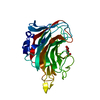 3a0nC  3gneSC S: Starting model for refinement C: citing same article ( |
|---|---|
| Similar structure data |


-Links
 PDBj
PDBj- Assembly
| Deposited unit | 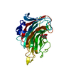
| ||||||||
|---|---|---|---|---|---|---|---|---|---|
| 1 |
| ||||||||
| Unit cell |
|
-Components
| #1: Protein | Mass: 28042.572 Da / Num. of mol.: 1 / Fragment: C-terminal domain, UNP residues 106-349 Source method: isolated from a genetically manipulated source Source: (gene. exp.) Chlorella virus / Strain: CVK-2 / Gene: vAL-1 / Plasmid: pET21b / Production host:  References: UniProt: Q9DTZ2, mannuronate-specific alginate lyase |
|---|---|
| #2: Sugar | ChemComp-BDP / 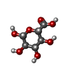  Type: D-saccharide, beta linking / Mass: 194.139 Da / Num. of mol.: 1 Type: D-saccharide, beta linking / Mass: 194.139 Da / Num. of mol.: 1Source method: isolated from a genetically manipulated source Formula: C6H10O7 |
| #3: Water | ChemComp-HOH /  Mass: 18.015 Da / Num. of mol.: 453 / Source method: isolated from a natural source / Formula: H2O Mass: 18.015 Da / Num. of mol.: 453 / Source method: isolated from a natural source / Formula: H2O |
| Nonpolymer details | SINCE O1 OF BDP IS MISSING IN THIS ENTRY, ALTERNATIV |
-Experimental details
-Experiment
| Experiment | Method: X-RAY DIFFRACTION / Number of used crystals: 1 |
|---|
- Sample preparation
| Crystal | Density Matthews: 2.58 Å3/Da / Density % sol: 52.32 % |
|---|---|
| Crystal grow | Temperature: 293 K / Method: vapor diffusion, sitting drop / pH: 7 Details: 12% PEG 3350, 0.2M ammonium formate, 0.1M hepes-NaOH pH7.0, VAPOR DIFFUSION, SITTING DROP, temperature 293K |
-Data collection
| Diffraction | Mean temperature: 100 K |
|---|---|
| Diffraction source | Source: SYNCHROTRON / Site: SPring-8  / Beamline: BL38B1 / Wavelength: 1 Å / Beamline: BL38B1 / Wavelength: 1 Å |
| Detector | Type: RIGAKU JUPITER 210 / Detector: CCD / Date: Jun 29, 2009 |
| Radiation | Monochromator: Fixed exit Si 111 double crystal monochromator Protocol: SINGLE WAVELENGTH / Monochromatic (M) / Laue (L): M / Scattering type: x-ray |
| Radiation wavelength | Wavelength: 1 Å / Relative weight: 1 |
| Reflection | Resolution: 1.66→26.32 Å / Num. all: 34464 / Num. obs: 34464 / % possible obs: 97.5 % / Observed criterion σ(F): 0 / Observed criterion σ(I): 0 / Redundancy: 5.4 % / Rmerge(I) obs: 0.04 / Net I/σ(I): 74.6 |
| Reflection shell | Resolution: 1.66→1.72 Å / Redundancy: 5.1 % / Rmerge(I) obs: 0.144 / Mean I/σ(I) obs: 16.5 / Num. unique all: 3330 / % possible all: 97.5 |
- Processing
| Software |
| |||||||||||||||||||||||||||||||||
|---|---|---|---|---|---|---|---|---|---|---|---|---|---|---|---|---|---|---|---|---|---|---|---|---|---|---|---|---|---|---|---|---|---|---|
| Refinement | Method to determine structure: AB INITIO Starting model: PDB ENTRY 3GNE Resolution: 1.66→10 Å / Num. parameters: 9879 / Num. restraintsaints: 8643 / Isotropic thermal model: Isotropic / Cross valid method: FREE R / σ(F): 0 / Stereochemistry target values: ENGH AND HUBER
| |||||||||||||||||||||||||||||||||
| Refine analyze | Num. disordered residues: 14 / Occupancy sum hydrogen: 0 / Occupancy sum non hydrogen: 2363.52 | |||||||||||||||||||||||||||||||||
| Refinement step | Cycle: LAST / Resolution: 1.66→10 Å
| |||||||||||||||||||||||||||||||||
| Refine LS restraints |
|
