 Movie
Movie Controller


+ Open data

- Basic information
| Entry | Database: PDB / ID: 1jud | ||||||
|---|---|---|---|---|---|---|---|
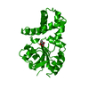
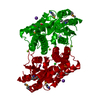
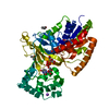
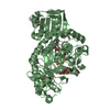
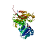
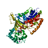
| Title | L-2-HALOACID DEHALOGENASE | ||||||
 Components Components | L-2-HALOACID DEHALOGENASE | ||||||
 Keywords Keywords | DEHALOGENASE / HYDROLASE | ||||||
| Function / homology |  Function and homology information Function and homology information | ||||||
| Biological species |  Pseudomonas sp. (bacteria) Pseudomonas sp. (bacteria) | ||||||
| Method |  X-RAY DIFFRACTION / MIR / Resolution: 2.5 Å X-RAY DIFFRACTION / MIR / Resolution: 2.5 Å | ||||||
 Authors Authors | Hisano, T. / Hata, Y. / Fujii, T. / Liu, J.-Q. / Kurihara, T. / Esaki, N. / Soda, K. | ||||||
 Citation Citation | Journal: J.Biol.Chem. / Year: 1996 Title: Crystal structure of L-2-haloacid dehalogenase from Pseudomonas sp. YL. An alpha/beta hydrolase structure that is different from the alpha/beta hydrolase fold. Authors: Hisano, T. / Hata, Y. / Fujii, T. / Liu, J.Q. / Kurihara, T. / Esaki, N. / Soda, K. #1: Journal: Proteins / Year: 1996Title: Crystallization of L-2-Haloacid Dehalogenase from Pseudomonas Sp. Yl Authors: Hisano, T. / Hata, Y. / Fujii, T. / Kurihara, T. / Esaki, N. / Soda, K. | ||||||
| History |
|
- Structure visualization
| Structure viewer | Molecule: MolmilJmol/JSmol |
|---|
- Downloads & links
-Download
| PDBx/mmCIF format | 1jud.cif.gz | 55.4 KB | Display | PDBx/mmCIF format |
|---|---|---|---|---|
| PDB format | pdb1jud.ent.gz | 40.4 KB | Display | PDB format |
| PDBx/mmJSON format | 1jud.json.gz | Tree view | PDBx/mmJSON format | |
| Others |  Other downloads Other downloads |
-Validation report
| Arichive directory | https://data.pdbj.org/pub/pdb/validation_reports/ju/1judftp://data.pdbj.org/pub/pdb/validation_reports/ju/1jud | HTTPS FTP |
|---|
-Related structure data
| Similar structure data |
|---|


-Links
 PDBj
PDBj

- Assembly
| Deposited unit | 
| ||||||||
|---|---|---|---|---|---|---|---|---|---|
| 1 | 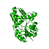
| ||||||||
| Unit cell |
|
-Components
| #1: Protein | Mass: 26207.588 Da / Num. of mol.: 1 Source method: isolated from a genetically manipulated source Source: (gene. exp.) Pseudomonas sp. (bacteria) / Strain: YL / Gene: L-DEX YL / Plasmid: PKK223-3 / Gene (production host): L-DEX YL / Production host: |
|---|---|
| #2: Water | ChemComp-HOH /  Mass: 18.015 Da / Num. of mol.: 62 / Source method: isolated from a natural source / Formula: H2O Mass: 18.015 Da / Num. of mol.: 62 / Source method: isolated from a natural source / Formula: H2O |
-Experimental details
-Experiment
| Experiment | Method: X-RAY DIFFRACTION / Number of used crystals: 1 |
|---|
- Sample preparation
| Crystal | Density Matthews: 2.37 Å3/Da / Density % sol: 48 % | |||||||||||||||||||||||||
|---|---|---|---|---|---|---|---|---|---|---|---|---|---|---|---|---|---|---|---|---|---|---|---|---|---|---|
| Crystal grow | pH: 5 / Details: pH 5.0 | |||||||||||||||||||||||||
| Crystal | *PLUS | |||||||||||||||||||||||||
| Crystal grow | *PLUS Temperature: 4 ℃ / Method: vapor diffusion | |||||||||||||||||||||||||
| Components of the solutions | *PLUS
|
-Data collection
| Diffraction source | Source: ROTATING ANODE / Type: RIGAKU RUH3R / Wavelength: 1.5418 |
|---|---|
| Detector | Type: RIGAKU / Detector: IMAGE PLATE / Date: Jan 7, 1995 |
| Radiation | Monochromator: GRAPHITE(002) / Monochromatic (M) / Laue (L): M / Scattering type: x-ray |
| Radiation wavelength | Wavelength: 1.5418 Å / Relative weight: 1 |
| Reflection | Resolution: 2.5→43 Å / Num. obs: 8124 / % possible obs: 93 % / Observed criterion σ(I): 1 / Redundancy: 3.9 % / Rmerge(I) obs: 0.0397 |
| Reflection shell | Resolution: 2.5→2.6 Å / Redundancy: 3.4 % / Rmerge(I) obs: 0.109 / % possible all: 90.3 |
| Reflection | *PLUS Num. measured all: 31020 |
- Processing
| Software |
| ||||||||||||||||||||||||||||||||||||||||||||||||||||||||||||
|---|---|---|---|---|---|---|---|---|---|---|---|---|---|---|---|---|---|---|---|---|---|---|---|---|---|---|---|---|---|---|---|---|---|---|---|---|---|---|---|---|---|---|---|---|---|---|---|---|---|---|---|---|---|---|---|---|---|---|---|---|---|
| Refinement | Method to determine structure: MIR / Resolution: 2.5→8 Å / σ(F): 2
| ||||||||||||||||||||||||||||||||||||||||||||||||||||||||||||
| Displacement parameters | Biso mean: 14.14 Å2 | ||||||||||||||||||||||||||||||||||||||||||||||||||||||||||||
| Refine analyze | Luzzati coordinate error obs: 0.25 Å | ||||||||||||||||||||||||||||||||||||||||||||||||||||||||||||
| Refinement step | Cycle: LAST / Resolution: 2.5→8 Å
| ||||||||||||||||||||||||||||||||||||||||||||||||||||||||||||
| Refine LS restraints |
| ||||||||||||||||||||||||||||||||||||||||||||||||||||||||||||
| Software | *PLUS Name: X-PLOR / Version: 3.1 / Classification: refinement | ||||||||||||||||||||||||||||||||||||||||||||||||||||||||||||
| Refinement | *PLUS | ||||||||||||||||||||||||||||||||||||||||||||||||||||||||||||
| Solvent computation | *PLUS | ||||||||||||||||||||||||||||||||||||||||||||||||||||||||||||
| Displacement parameters | *PLUS Biso mean: 14.143 Å2 | ||||||||||||||||||||||||||||||||||||||||||||||||||||||||||||
| Refine LS restraints | *PLUS
|
