 Movie
Movie Controller

[English] 日本語
 Yorodumi
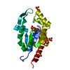
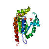
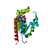
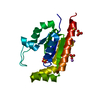
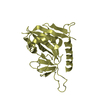
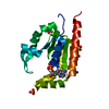
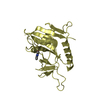
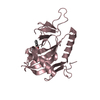
- PDB-3iij: The structure of hCINAP-ADP complex at 1.76 angstroms resolution. -
+ Open data

- Basic information
| Entry | Database: PDB / ID: 3iij | ||||||
|---|---|---|---|---|---|---|---|
| Title | The structure of hCINAP-ADP complex at 1.76 angstroms resolution. | ||||||
 Components Components | Coilin-interacting nuclear ATPase protein | ||||||
 Keywords Keywords | PROTEIN BINDING / TRANSFERASE / Alpha and beta proteins (a/b) / phosphotransferase | ||||||
| Function / homology |  Function and homology information Function and homology informationnucleoside monophosphate kinase activity / adenylate kinase / adenylate kinase activity / nucleobase-containing small molecule interconversion / Interconversion of nucleotide di- and triphosphates / Cajal body / rRNA processing / ribosomal small subunit biogenesis / nuclear speck / phosphorylation ...nucleoside monophosphate kinase activity / adenylate kinase / adenylate kinase activity / nucleobase-containing small molecule interconversion / Interconversion of nucleotide di- and triphosphates / Cajal body / rRNA processing / ribosomal small subunit biogenesis / nuclear speck / phosphorylation / centrosome / ATP hydrolysis activity / nucleoplasm / ATP binding / membrane / nucleus / cytoplasm Similarity search - Function | ||||||
| Biological species |  Homo sapiens (human) Homo sapiens (human) | ||||||
| Method |  X-RAY DIFFRACTION / SYNCHROTRON / MOLECULAR REPLACEMENT / Resolution: 1.76 Å X-RAY DIFFRACTION / SYNCHROTRON / MOLECULAR REPLACEMENT / Resolution: 1.76 Å | ||||||
 Authors Authors | Zographos, S.E. / Drakou, C.E. / Leonidas, D.D. | ||||||
 Citation Citation | Journal: Proteins / Year: 2012 Title: hCINAP is an atypical mammalian nuclear adenylate kinase with an ATPase motif: Structural and functional studies. Authors: Drakou, C.E. / Malekkou, A. / Hayes, J.M. / Lederer, C.W. / Leonidas, D.D. / Oikonomakos, N.G. / Lamond, A.I. / Santama, N. / Zographos, S.E. | ||||||
| History |
|
- Structure visualization
| Structure viewer | Molecule: MolmilJmol/JSmol |
|---|
- Downloads & links
-Download
| PDBx/mmCIF format | 3iij.cif.gz | 58 KB | Display | PDBx/mmCIF format |
|---|---|---|---|---|
| PDB format | pdb3iij.ent.gz | 41.9 KB | Display | PDB format |
| PDBx/mmJSON format | 3iij.json.gz | Tree view | PDBx/mmJSON format | |
| Others |  Other downloads Other downloads |
-Validation report
| Summary document | 3iij_validation.pdf.gz | 814.8 KB | Display | wwPDB validaton report |
|---|---|---|---|---|
| Full document | 3iij_full_validation.pdf.gz | 817.5 KB | Display | |
| Data in XML | 3iij_validation.xml.gz | 11.3 KB | Display | |
| Data in CIF | 3iij_validation.cif.gz | 16.1 KB | Display | |
| Arichive directory | https://data.pdbj.org/pub/pdb/validation_reports/ii/3iijftp://data.pdbj.org/pub/pdb/validation_reports/ii/3iij | HTTPS FTP |
-Related structure data
| Related structure data |  3iikC  3iilC  3iimC  1rkbS S: Starting model for refinement C: citing same article ( |
|---|---|
| Similar structure data |


-Links
 PDBj
PDBj

- Assembly
| Deposited unit | 
| ||||||||
|---|---|---|---|---|---|---|---|---|---|
| 1 |
| ||||||||
| Unit cell |
|
-Components
| #1: Protein | Mass: 20867.262 Da / Num. of mol.: 1 Source method: isolated from a genetically manipulated source Source: (gene. exp.) Homo sapiens (human) / Gene: CINAP, TAF9, hCG_37060 / Plasmid: pGEX-6P-3 / Production host:  References: UniProt: Q5F2S9, UniProt: Q9Y3D8*PLUS, adenylate kinase | ||
|---|---|---|---|
| #2: Chemical | ChemComp-ADP /   Mass: 427.201 Da / Num. of mol.: 1 / Source method: obtained synthetically / Formula: C10H15N5O10P2 / Comment: ADP, energy-carrying molecule*YM Mass: 427.201 Da / Num. of mol.: 1 / Source method: obtained synthetically / Formula: C10H15N5O10P2 / Comment: ADP, energy-carrying molecule*YM | ||
| #3: Chemical | ChemComp-SO4 /   Mass: 96.063 Da / Num. of mol.: 4 / Source method: obtained synthetically / Formula: SO4 Mass: 96.063 Da / Num. of mol.: 4 / Source method: obtained synthetically / Formula: SO4#4: Water | ChemComp-HOH / |  Mass: 18.015 Da / Num. of mol.: 190 / Source method: isolated from a natural source / Formula: H2O Mass: 18.015 Da / Num. of mol.: 190 / Source method: isolated from a natural source / Formula: H2O |
-Experimental details
-Experiment
| Experiment | Method: X-RAY DIFFRACTION / Number of used crystals: 1 |
|---|
- Sample preparation
| Crystal | Density Matthews: 3.93 Å3/Da / Density % sol: 68.72 % |
|---|---|
| Crystal grow | Temperature: 293 K / Method: vapor diffusion, hanging drop / pH: 7.5 Details: 0.1 M HEPES pH 7.5, 1.5 M Li2SO4, 0.2 M NaCl, 0.5 mM DTT, 25 mM MgCl2, 2 mM ADP, VAPOR DIFFUSION, HANGING DROP, temperature 293K |
-Data collection
| Diffraction | Mean temperature: 100 K |
|---|---|
| Diffraction source | Source: SYNCHROTRON / Site: SRS  / Beamline: PX10.1 / Wavelength: 1.11665 Å / Beamline: PX10.1 / Wavelength: 1.11665 Å |
| Detector | Type: MARMOSAIC 225 mm CCD / Detector: CCD / Date: Jan 1, 2007 / Details: Rh Coated mirrors |
| Radiation | Monochromator: double crystal monochromator with sagittal focussing Protocol: SINGLE WAVELENGTH / Monochromatic (M) / Laue (L): M / Scattering type: x-ray |
| Radiation wavelength | Wavelength: 1.11665 Å / Relative weight: 1 |
| Reflection | Resolution: 1.76→30 Å / Num. all: 32104 / Num. obs: 31610 / % possible obs: 98.1 % / Observed criterion σ(I): 2 / Redundancy: 4.9 % / Biso Wilson estimate: 32.8 Å2 / Rsym value: 0.044 / Net I/σ(I): 17.92 |
| Reflection shell | Resolution: 1.76→1.79 Å / Redundancy: 4.3 % / Mean I/σ(I) obs: 3.41 / Rsym value: 0.499 / % possible all: 88.4 |
- Processing
| Software |
| |||||||||||||||||||||||||||||||||||||||||||||||||||||||||||||||||||||||||||||||||||||||||||||||||||||||||||||||||||||||||||||||||||||||||||||||||||||||||||||||||||||||||||||||||||||||||||||||||||||||||||||||||||||||||||||||||||||||||||||||||||||||||||||||||||||||||||||||||||||||||||||||||||||||||||||||||||||||||||||||||||||
|---|---|---|---|---|---|---|---|---|---|---|---|---|---|---|---|---|---|---|---|---|---|---|---|---|---|---|---|---|---|---|---|---|---|---|---|---|---|---|---|---|---|---|---|---|---|---|---|---|---|---|---|---|---|---|---|---|---|---|---|---|---|---|---|---|---|---|---|---|---|---|---|---|---|---|---|---|---|---|---|---|---|---|---|---|---|---|---|---|---|---|---|---|---|---|---|---|---|---|---|---|---|---|---|---|---|---|---|---|---|---|---|---|---|---|---|---|---|---|---|---|---|---|---|---|---|---|---|---|---|---|---|---|---|---|---|---|---|---|---|---|---|---|---|---|---|---|---|---|---|---|---|---|---|---|---|---|---|---|---|---|---|---|---|---|---|---|---|---|---|---|---|---|---|---|---|---|---|---|---|---|---|---|---|---|---|---|---|---|---|---|---|---|---|---|---|---|---|---|---|---|---|---|---|---|---|---|---|---|---|---|---|---|---|---|---|---|---|---|---|---|---|---|---|---|---|---|---|---|---|---|---|---|---|---|---|---|---|---|---|---|---|---|---|---|---|---|---|---|---|---|---|---|---|---|---|---|---|---|---|---|---|---|---|---|---|---|---|---|---|---|---|---|---|---|---|---|---|---|---|---|---|---|---|---|---|---|---|---|---|---|---|---|---|---|---|---|---|---|---|---|---|---|---|---|---|---|---|---|---|---|---|---|---|---|---|---|---|---|---|---|---|---|---|---|---|---|
| Refinement | Method to determine structure: MOLECULAR REPLACEMENT Starting model: 1RKB Resolution: 1.76→28.3 Å / Cor.coef. Fo:Fc: 0.963 / Cor.coef. Fo:Fc free: 0.955 / SU B: 3.051 / SU ML: 0.049 / TLS residual ADP flag: LIKELY RESIDUAL / Cross valid method: THROUGHOUT / σ(F): 2 / ESU R: 0.089 / ESU R Free: 0.084 / Stereochemistry target values: MAXIMUM LIKELIHOOD / Details: HYDROGENS HAVE BEEN ADDED IN THE RIDING POSITIONS
| |||||||||||||||||||||||||||||||||||||||||||||||||||||||||||||||||||||||||||||||||||||||||||||||||||||||||||||||||||||||||||||||||||||||||||||||||||||||||||||||||||||||||||||||||||||||||||||||||||||||||||||||||||||||||||||||||||||||||||||||||||||||||||||||||||||||||||||||||||||||||||||||||||||||||||||||||||||||||||||||||||||
| Solvent computation | Ion probe radii: 0.8 Å / Shrinkage radii: 0.8 Å / VDW probe radii: 1.4 Å / Solvent model: MASK | |||||||||||||||||||||||||||||||||||||||||||||||||||||||||||||||||||||||||||||||||||||||||||||||||||||||||||||||||||||||||||||||||||||||||||||||||||||||||||||||||||||||||||||||||||||||||||||||||||||||||||||||||||||||||||||||||||||||||||||||||||||||||||||||||||||||||||||||||||||||||||||||||||||||||||||||||||||||||||||||||||||
| Displacement parameters | Biso mean: 21.63 Å2
| |||||||||||||||||||||||||||||||||||||||||||||||||||||||||||||||||||||||||||||||||||||||||||||||||||||||||||||||||||||||||||||||||||||||||||||||||||||||||||||||||||||||||||||||||||||||||||||||||||||||||||||||||||||||||||||||||||||||||||||||||||||||||||||||||||||||||||||||||||||||||||||||||||||||||||||||||||||||||||||||||||||
| Refinement step | Cycle: LAST / Resolution: 1.76→28.3 Å
| |||||||||||||||||||||||||||||||||||||||||||||||||||||||||||||||||||||||||||||||||||||||||||||||||||||||||||||||||||||||||||||||||||||||||||||||||||||||||||||||||||||||||||||||||||||||||||||||||||||||||||||||||||||||||||||||||||||||||||||||||||||||||||||||||||||||||||||||||||||||||||||||||||||||||||||||||||||||||||||||||||||
| Refine LS restraints |
| |||||||||||||||||||||||||||||||||||||||||||||||||||||||||||||||||||||||||||||||||||||||||||||||||||||||||||||||||||||||||||||||||||||||||||||||||||||||||||||||||||||||||||||||||||||||||||||||||||||||||||||||||||||||||||||||||||||||||||||||||||||||||||||||||||||||||||||||||||||||||||||||||||||||||||||||||||||||||||||||||||||
| LS refinement shell | Resolution: 1.76→1.809 Å / Total num. of bins used: 20
| |||||||||||||||||||||||||||||||||||||||||||||||||||||||||||||||||||||||||||||||||||||||||||||||||||||||||||||||||||||||||||||||||||||||||||||||||||||||||||||||||||||||||||||||||||||||||||||||||||||||||||||||||||||||||||||||||||||||||||||||||||||||||||||||||||||||||||||||||||||||||||||||||||||||||||||||||||||||||||||||||||||
| Refinement TLS params. | Method: refined / Refine-ID: X-RAY DIFFRACTION
| |||||||||||||||||||||||||||||||||||||||||||||||||||||||||||||||||||||||||||||||||||||||||||||||||||||||||||||||||||||||||||||||||||||||||||||||||||||||||||||||||||||||||||||||||||||||||||||||||||||||||||||||||||||||||||||||||||||||||||||||||||||||||||||||||||||||||||||||||||||||||||||||||||||||||||||||||||||||||||||||||||||
| Refinement TLS group |
|
