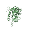
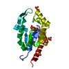
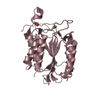
Entry Database : PDB / ID : 5jzvTitle The structure of D77G hCINAP-ADP Adenylate kinase isoenzyme 6 Keywords / / Function / homology Function Domain/homology Component
/ / / / / / / / / / / / / / / / / / / / / / / / / / / / / / Biological species Homo sapiens (human)Method / / / Resolution : 2.07 Å Authors Liu, Y. / Yang, Z. / Yang, Y. / Cai, X. / Zheng, X. Funding support Organization Grant number Country National Science Foundation of China 31470754 Doctoral Fund of the Ministry of Education of China 20130001130003 Beijing Natural Science Foundation Grant 5152012
Journal : Nat Commun / Year : 2016Title : The ATPase hCINAP regulates 18S rRNA processing and is essential for embryogenesis and tumour growth.Authors : Bai, D. / Zhang, J. / Li, T. / Hang, R. / Liu, Y. / Tian, Y. / Huang, D. / Qu, L. / Cao, X. / Ji, J. / Zheng, X. History Deposition May 17, 2016 Deposition site / Processing site Revision 1.0 Aug 10, 2016 Provider / Type Revision 1.1 Dec 6, 2017 Group / Derived calculationsCategory / citation_author / pdbx_struct_oper_listItem _citation.journal_volume / _citation.page_first ... _citation.journal_volume / _citation.page_first / _citation.page_last / _citation.pdbx_database_id_DOI / _citation.pdbx_database_id_PubMed / _citation.title / _pdbx_struct_oper_list.symmetry_operation Revision 1.2 Nov 8, 2023 Group / Database references / Refinement descriptionCategory chem_comp_atom / chem_comp_bond ... chem_comp_atom / chem_comp_bond / database_2 / pdbx_initial_refinement_model Item / _database_2.pdbx_database_accession
Show all Show less
 Movie
Movie Controller


 Open data
 Basic information
 Components
 Keywords
 Function and homology information
 Homo sapiens (human)
 X-RAY DIFFRACTION /
  Authors
 China, 3items
  Citation
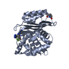
 Structure visualization
 Downloads & links
 Other downloads


 PDBj
PDBj

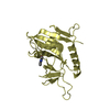
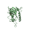
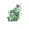
 Assembly
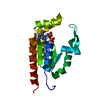


 Mass: 427.201 Da / Num. of mol.: 1 / Source method: obtained synthetically / Formula: C10H15N5O10P2 / Comment: ADP, energy-carrying molecule*YM
 Mass: 18.015 Da / Num. of mol.: 171 / Source method: isolated from a natural source / Formula: H2O
 Sample preparation
 Processing