 Movie
Movie Controller

[English] 日本語
 Yorodumi
- PDB-3ihq: Crystal Structure of Reduced C10S Spx in Complex with the Alpha C... -
+ Open data

- Basic information
| Entry | Database: PDB / ID: 3ihq | ||||||
|---|---|---|---|---|---|---|---|
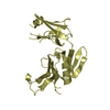
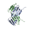
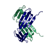
| Title | Crystal Structure of Reduced C10S Spx in Complex with the Alpha C-terminal Domain of RNA Polymeras | ||||||
 Components Components |
| ||||||
 Keywords Keywords | Transcription/Transferase / transcription regulation / oxidative stress / Spx / RNA Polymerase / Cytoplasm / Disulfide bond / Redox-active center / Stress response / Transcription / DNA-directed RNA polymerase / Nucleotidyltransferase / Transferase / Transcription-Transferase COMPLEX | ||||||
| Function / homology |  Function and homology information Function and homology informationDNA-directed RNA polymerase complex / DNA-directed RNA polymerase / DNA-directed RNA polymerase activity / protein dimerization activity / negative regulation of DNA-templated transcription / DNA-templated transcription / DNA binding / cytoplasm Similarity search - Function | ||||||
| Biological species |  | ||||||
| Method |  X-RAY DIFFRACTION / SYNCHROTRON / MOLECULAR REPLACEMENT / Resolution: 1.9 Å X-RAY DIFFRACTION / SYNCHROTRON / MOLECULAR REPLACEMENT / Resolution: 1.9 Å | ||||||
 Authors Authors | Newberry, K.J. / Brennan, R.G. | ||||||
 Citation Citation | Journal: Plos One / Year: 2010 Title: Promoter recognition by a complex of Spx and the C-terminal domain of the RNA polymerase alpha subunit. Authors: Nakano, M.M. / Lin, A. / Zuber, C.S. / Newberry, K.J. / Brennan, R.G. / Zuber, P. #1: Journal: Proc.Natl.Acad.Sci.USA / Year: 2005Title: Crystal structure of the Bacillus subtilis anti-alpha, global transcriptional regulator, Spx, in complex with the {alpha} C-terminal domain of RNA polymerase Authors: Newberry, K.J. / Nakano, S. / Zuber, P. / Brennan, R.G. | ||||||
| History |
|
- Structure visualization
| Structure viewer | Molecule: MolmilJmol/JSmol |
|---|
- Downloads & links
-Download
| PDBx/mmCIF format | 3ihq.cif.gz | 52.3 KB | Display | PDBx/mmCIF format |
|---|---|---|---|---|
| PDB format | pdb3ihq.ent.gz | 36.1 KB | Display | PDB format |
| PDBx/mmJSON format | 3ihq.json.gz | Tree view | PDBx/mmJSON format | |
| Others |  Other downloads Other downloads |
-Validation report
| Arichive directory | https://data.pdbj.org/pub/pdb/validation_reports/ih/3ihqftp://data.pdbj.org/pub/pdb/validation_reports/ih/3ihq | HTTPS FTP |
|---|
-Related structure data
| Related structure data | 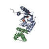 1z3eS S: Starting model for refinement |
|---|---|
| Similar structure data |


-Links
 PDBj
PDBj


- Assembly
| Deposited unit | 
| ||||||||
|---|---|---|---|---|---|---|---|---|---|
| 1 |
| ||||||||
| Unit cell |
|
-Components
| #1: Protein | Mass: 15673.976 Da / Num. of mol.: 1 / Mutation: C10S Source method: isolated from a genetically manipulated source Source: (gene. exp.) |
|---|---|
| #2: Protein | Mass: 8534.862 Da / Num. of mol.: 1 / Fragment: UNP residues 245-314 Source method: isolated from a genetically manipulated source Source: (gene. exp.) |
| #3: Chemical | ChemComp-IMD /   Mass: 69.085 Da / Num. of mol.: 1 / Source method: obtained synthetically / Formula: C3H5N2 Mass: 69.085 Da / Num. of mol.: 1 / Source method: obtained synthetically / Formula: C3H5N2 |
| #4: Water | ChemComp-HOH /  Mass: 18.015 Da / Num. of mol.: 39 / Source method: isolated from a natural source / Formula: H2O Mass: 18.015 Da / Num. of mol.: 39 / Source method: isolated from a natural source / Formula: H2O |
-Experimental details
-Experiment
| Experiment | Method: X-RAY DIFFRACTION / Number of used crystals: 1 |
|---|
- Sample preparation
| Crystal | Density Matthews: 1.85 Å3/Da / Density % sol: 33.47 % |
|---|---|
| Crystal grow | Temperature: 273 K / Method: vapor diffusion, hanging drop / pH: 5.3 Details: 25-30% PEG 4000, 0.1 M sodium citrate, 0.1 M magnesium chloride, pH 5.3, VAPOR DIFFUSION, HANGING DROP, temperature 273K |
-Data collection
| Diffraction | Mean temperature: 100 K |
|---|---|
| Diffraction source | Source: SYNCHROTRON / Site: ALS  / Beamline: 8.3.1 / Wavelength: 1 Å / Beamline: 8.3.1 / Wavelength: 1 Å |
| Detector | Type: ADSC QUANTUM 315r / Detector: CCD / Date: Jun 22, 2006 / Details: Double Crystal Si(111) |
| Radiation | Protocol: SINGLE WAVELENGTH / Monochromatic (M) / Laue (L): M / Scattering type: x-ray |
| Radiation wavelength | Wavelength: 1 Å / Relative weight: 1 |
| Reflection | Resolution: 1.9→48.33 Å / Num. obs: 12912 / % possible obs: 95.1 % / Observed criterion σ(F): 2 / Redundancy: 3.5 % / Biso Wilson estimate: 27 Å2 / Rmerge(I) obs: 0.059 / Rsym value: 0.059 / Net I/σ(I): 7.7 |
| Reflection shell | Resolution: 1.9→2 Å / Redundancy: 3.4 % / Rmerge(I) obs: 0.23 / Mean I/σ(I) obs: 2.9 / Num. unique all: 12912 / Rsym value: 0.23 / % possible all: 94.1 |
- Processing
| Software |
| |||||||||||||||||||||||||
|---|---|---|---|---|---|---|---|---|---|---|---|---|---|---|---|---|---|---|---|---|---|---|---|---|---|---|
| Refinement | Method to determine structure: MOLECULAR REPLACEMENT Starting model: PDB entry 1Z3E Resolution: 1.9→48.33 Å / Isotropic thermal model: isotropic / Cross valid method: THROUGHOUT / σ(F): 2 / Stereochemistry target values: Engh & Huber
| |||||||||||||||||||||||||
| Displacement parameters | Biso mean: 38.4 Å2 | |||||||||||||||||||||||||
| Refine analyze |
| |||||||||||||||||||||||||
| Refinement step | Cycle: LAST / Resolution: 1.9→48.33 Å
| |||||||||||||||||||||||||
| LS refinement shell | Resolution: 1.9→1.97 Å
|
