 Movie
Movie Controller


+ Open data

- Basic information
| Entry | Database: PDB / ID: 3iep | ||||||
|---|---|---|---|---|---|---|---|
| Title | Firefly luciferase apo structure (P41 form) | ||||||
 Components Components | Luciferin 4-monooxygenase | ||||||
 Keywords Keywords | OXIDOREDUCTASE / MONOOXYGENASE / PHOTOPROTEIN / LUMINESCENCE / ATP-binding / Magnesium / Metal-binding / Nucleotide-binding / Peroxisome | ||||||
| Function / homology |  Function and homology information Function and homology informationPhotinus-luciferin 4-monooxygenase (ATP-hydrolyzing) activity / firefly luciferase / CoA-ligase activity / bioluminescence / peroxisome / protein-folding chaperone binding / ATP binding / metal ion binding Similarity search - Function | ||||||
| Biological species |  Photinus pyralis (common eastern firefly) Photinus pyralis (common eastern firefly) | ||||||
| Method |  X-RAY DIFFRACTION / SYNCHROTRON / MOLECULAR REPLACEMENT / molecular replacement / Resolution: 2.1 Å X-RAY DIFFRACTION / SYNCHROTRON / MOLECULAR REPLACEMENT / molecular replacement / Resolution: 2.1 Å | ||||||
 Authors Authors | Lovell, S. / Battaile, K.P. / Auld, D.S. / Thorne, N. / Lea, W.A. / Maloney, D.J. / Shen, M. / Raj, G. / Thomas, C.J. / Simeonov, A. ...Lovell, S. / Battaile, K.P. / Auld, D.S. / Thorne, N. / Lea, W.A. / Maloney, D.J. / Shen, M. / Raj, G. / Thomas, C.J. / Simeonov, A. / Hanzlik, R.P. / Inglese, J. | ||||||
 Citation Citation | Journal: Proc.Natl.Acad.Sci.USA / Year: 2010 Title: Molecular basis for the high-affinity binding and stabilization of firefly luciferase by PTC124. Authors: Auld, D.S. / Lovell, S. / Thorne, N. / Lea, W.A. / Maloney, D.J. / Shen, M. / Rai, G. / Battaile, K.P. / Thomas, C.J. / Simeonov, A. / Hanzlik, R.P. / Inglese, J. | ||||||
| History |
|
- Structure visualization
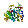
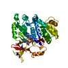
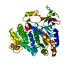
| Structure viewer | Molecule: MolmilJmol/JSmol |
|---|
- Downloads & links
-Download
| PDBx/mmCIF format | 3iep.cif.gz | 101.6 KB | Display | PDBx/mmCIF format |
|---|---|---|---|---|
| PDB format | pdb3iep.ent.gz | 75.8 KB | Display | PDB format |
| PDBx/mmJSON format | 3iep.json.gz | Tree view | PDBx/mmJSON format | |
| Others |  Other downloads Other downloads |
-Validation report
| Arichive directory | https://data.pdbj.org/pub/pdb/validation_reports/ie/3iepftp://data.pdbj.org/pub/pdb/validation_reports/ie/3iep | HTTPS FTP |
|---|
-Related structure data


-Links
 PDBj
PDBj
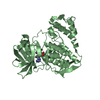
- Assembly
| Deposited unit | 
| ||||||||
|---|---|---|---|---|---|---|---|---|---|
| 1 |
| ||||||||
| Unit cell |
|
-Components
| #1: Protein | Mass: 60918.086 Da / Num. of mol.: 1 / Source method: isolated from a natural source / Source: (natural) Photinus pyralis (common eastern firefly) / References: UniProt: P08659, firefly luciferase |
|---|---|
| #2: Water | ChemComp-HOH /  Mass: 18.015 Da / Num. of mol.: 199 / Source method: isolated from a natural source / Formula: H2O Mass: 18.015 Da / Num. of mol.: 199 / Source method: isolated from a natural source / Formula: H2O |
-Experimental details
-Experiment
| Experiment | Method: X-RAY DIFFRACTION / Number of used crystals: 1 |
|---|
- Sample preparation
| Crystal | Density Matthews: 2.85 Å3/Da / Density % sol: 56.84 % |
|---|---|
| Crystal grow | Temperature: 277 K / Method: vapor diffusion / pH: 8.5 Details: 30% PEG 1500, 8% MPD, 0.1M Tris-HCl, pH 8.5, VAPOR DIFFUSION, temperature 277K |
-Data collection
| Diffraction | Mean temperature: 100 K |
|---|---|
| Diffraction source | Source: SYNCHROTRON / Site: APS  / Beamline: 17-BM / Wavelength: 1 Å / Beamline: 17-BM / Wavelength: 1 Å |
| Detector | Type: ADSC QUANTUM 210 / Detector: CCD / Date: Mar 13, 2009 |
| Diffraction measurement | Details: 1.00 degrees, 12.0 sec, detector distance 150.00 mm |
| Radiation | Monochromator: Si(111) double crystal / Protocol: SINGLE WAVELENGTH / Scattering type: x-ray |
| Radiation wavelength | Wavelength: 1 Å / Relative weight: 1 |
| Reflection | Av R equivalents: 0.132 / Number: 202033 |
| Reflection | Resolution: 2.1→50 Å / Num. obs: 39872 / % possible obs: 99.9 % / Observed criterion σ(I): -3 / Redundancy: 5.1 % / Rmerge(I) obs: 0.132 / Rsym value: 0.132 / Net I/σ(I): 11.343 |
| Reflection shell | Resolution: 2.1→2.18 Å / Redundancy: 5 % / Rmerge(I) obs: 0.518 / Mean I/σ(I) obs: 2.748 / Rsym value: 0.518 / % possible all: 100 |
| Cell measurement | Reflection used: 202033 |
-Phasing
| Phasing | Method: molecular replacement | |||||||||
|---|---|---|---|---|---|---|---|---|---|---|
| Phasing MR |
|
- Processing
| Software |
| |||||||||||||||||||||||||||||||||||||||||||||||||||||||||||||||||||||||||||||||||||||||||||||||||||||||||||||||||||||||||||||||||||||||||||||||||||
|---|---|---|---|---|---|---|---|---|---|---|---|---|---|---|---|---|---|---|---|---|---|---|---|---|---|---|---|---|---|---|---|---|---|---|---|---|---|---|---|---|---|---|---|---|---|---|---|---|---|---|---|---|---|---|---|---|---|---|---|---|---|---|---|---|---|---|---|---|---|---|---|---|---|---|---|---|---|---|---|---|---|---|---|---|---|---|---|---|---|---|---|---|---|---|---|---|---|---|---|---|---|---|---|---|---|---|---|---|---|---|---|---|---|---|---|---|---|---|---|---|---|---|---|---|---|---|---|---|---|---|---|---|---|---|---|---|---|---|---|---|---|---|---|---|---|---|---|---|
| Refinement | Method to determine structure: MOLECULAR REPLACEMENT / Resolution: 2.1→50 Å / Cor.coef. Fo:Fc: 0.939 / Cor.coef. Fo:Fc free: 0.915 / WRfactor Rfree: 0.196 / WRfactor Rwork: 0.169 / Occupancy max: 1 / Occupancy min: 0.5 / SU B: 3.524 / SU ML: 0.094 / Cross valid method: THROUGHOUT / σ(F): 0 / ESU R: 0.155 / ESU R Free: 0.145 / Stereochemistry target values: MAXIMUM LIKELIHOOD Details: 1. HYDROGENS HAVE BEEN ADDED IN THE RIDING POSITIONS. 2. U VALUES: REFINED INDIVIDUALLY.
| |||||||||||||||||||||||||||||||||||||||||||||||||||||||||||||||||||||||||||||||||||||||||||||||||||||||||||||||||||||||||||||||||||||||||||||||||||
| Solvent computation | Ion probe radii: 0.8 Å / Shrinkage radii: 0.8 Å / VDW probe radii: 1.4 Å / Solvent model: BABINET MODEL WITH MASK | |||||||||||||||||||||||||||||||||||||||||||||||||||||||||||||||||||||||||||||||||||||||||||||||||||||||||||||||||||||||||||||||||||||||||||||||||||
| Displacement parameters | Biso max: 63.45 Å2 / Biso mean: 31.12 Å2 / Biso min: 19.22 Å2
| |||||||||||||||||||||||||||||||||||||||||||||||||||||||||||||||||||||||||||||||||||||||||||||||||||||||||||||||||||||||||||||||||||||||||||||||||||
| Refinement step | Cycle: LAST / Resolution: 2.1→50 Å
| |||||||||||||||||||||||||||||||||||||||||||||||||||||||||||||||||||||||||||||||||||||||||||||||||||||||||||||||||||||||||||||||||||||||||||||||||||
| Refine LS restraints |
| |||||||||||||||||||||||||||||||||||||||||||||||||||||||||||||||||||||||||||||||||||||||||||||||||||||||||||||||||||||||||||||||||||||||||||||||||||
| LS refinement shell | Refine-ID: X-RAY DIFFRACTION / Total num. of bins used: 20
|
