 Movie
Movie Controller

[English] 日本語
 Yorodumi
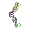
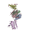
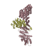
- PDB-3idx: Crystal structure of HIV-gp120 core in complex with CD4-binding s... -
+ Open data

- Basic information
| Entry | Database: PDB / ID: 3idx | ||||||
|---|---|---|---|---|---|---|---|
| Title | Crystal structure of HIV-gp120 core in complex with CD4-binding site antibody b13, space group C222 | ||||||
 Components Components |
| ||||||
 Keywords Keywords | IMMUNE SYSTEM / HIV-1 / antibody / gp120 / b13 / Envelope glycan protein / CD4-binding site / AIDS / Apoptosis / Cell membrane / Cleavage on pair of basic residues / Disulfide bond / Envelope protein / Fusion protein / Host-virus interaction / Membrane / Transmembrane / Viral immunoevasion / Virion | ||||||
| Function / homology |  Function and homology information Function and homology informationSynthesis and processing of ENV and VPU / symbiont-mediated evasion of host immune response / positive regulation of establishment of T cell polarity / Alpha-defensins / Dectin-2 family / Binding and entry of HIV virion / positive regulation of plasma membrane raft polarization / positive regulation of receptor clustering / host cell endosome membrane / actin filament organization ...Synthesis and processing of ENV and VPU / symbiont-mediated evasion of host immune response / positive regulation of establishment of T cell polarity / Alpha-defensins / Dectin-2 family / Binding and entry of HIV virion / positive regulation of plasma membrane raft polarization / positive regulation of receptor clustering / host cell endosome membrane / actin filament organization / Assembly Of The HIV Virion / Budding and maturation of HIV virion / clathrin-dependent endocytosis of virus by host cell / viral protein processing / receptor ligand activity / fusion of virus membrane with host plasma membrane / fusion of virus membrane with host endosome membrane / viral envelope / symbiont entry into host cell / virion attachment to host cell / host cell plasma membrane / virion membrane / structural molecule activity / membrane Similarity search - Function | ||||||
| Biological species |   Human immunodeficiency virus 1 Human immunodeficiency virus 1 Homo sapiens (human) Homo sapiens (human) | ||||||
| Method |  X-RAY DIFFRACTION / SYNCHROTRON / MOLECULAR REPLACEMENT / Resolution: 2.5 Å X-RAY DIFFRACTION / SYNCHROTRON / MOLECULAR REPLACEMENT / Resolution: 2.5 Å | ||||||
 Authors Authors | Chen, L. / Kwon, Y.D. / Zhou, T. / Wu, X. / O'Dell, S. / Cavacini, L. / Hessell, A.J. / Pancera, M. / Tang, M. / Xu, L. ...Chen, L. / Kwon, Y.D. / Zhou, T. / Wu, X. / O'Dell, S. / Cavacini, L. / Hessell, A.J. / Pancera, M. / Tang, M. / Xu, L. / Yang, Z.Y. / Zhang, M.Y. / Arthos, J. / Burton, D.R. / Dimitrov, D.S. / Nabel, G.J. / Posner, M. / Sodroski, J. / Wyatt, R. / Mascola, J.R. / Kwong, P.D. | ||||||
 Citation Citation | Journal: Science / Year: 2009 Title: Structural basis of immune evasion at the site of CD4 attachment on HIV-1 gp120. Authors: Chen, L. / Do Kwon, Y. / Zhou, T. / Wu, X. / O'Dell, S. / Cavacini, L. / Hessell, A.J. / Pancera, M. / Tang, M. / Xu, L. / Yang, Z.Y. / Zhang, M.Y. / Arthos, J. / Burton, D.R. / Dimitrov, D. ...Authors: Chen, L. / Do Kwon, Y. / Zhou, T. / Wu, X. / O'Dell, S. / Cavacini, L. / Hessell, A.J. / Pancera, M. / Tang, M. / Xu, L. / Yang, Z.Y. / Zhang, M.Y. / Arthos, J. / Burton, D.R. / Dimitrov, D.S. / Nabel, G.J. / Posner, M.R. / Sodroski, J. / Wyatt, R. / Mascola, J.R. / Kwong, P.D. | ||||||
| History |
|
- Structure visualization
| Structure viewer | Molecule: MolmilJmol/JSmol |
|---|
- Downloads & links
-Download
| PDBx/mmCIF format | 3idx.cif.gz | 322.3 KB | Display | PDBx/mmCIF format |
|---|---|---|---|---|
| PDB format | pdb3idx.ent.gz | 264.7 KB | Display | PDB format |
| PDBx/mmJSON format | 3idx.json.gz | Tree view | PDBx/mmJSON format | |
| Others |  Other downloads Other downloads |
-Validation report
| Arichive directory | https://data.pdbj.org/pub/pdb/validation_reports/id/3idxftp://data.pdbj.org/pub/pdb/validation_reports/id/3idx | HTTPS FTP |
|---|
-Related structure data
| Related structure data | 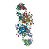 3hi1C 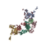 3idyC  1bbjS C: citing same article ( S: Starting model for refinement |
|---|---|
| Similar structure data |


-Links
 PDBj
PDBj


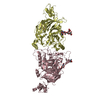
- Assembly
| Deposited unit | 
| ||||||||
|---|---|---|---|---|---|---|---|---|---|
| 1 |
| ||||||||
| Unit cell |
| ||||||||
| Components on special symmetry positions |
| ||||||||
| Details | Antigen/antibody complex: the biological assembly is chain G in complex with chain H and L |
-Components
-Antibody , 2 types, 2 molecules HL
| #2: Antibody | Mass: 24810.910 Da / Num. of mol.: 1 Source method: isolated from a genetically manipulated source Source: (gene. exp.) Homo sapiens (human) / Plasmid: pcDNA3.1(-) / Cell line (production host): HEK293F / Production host: Homo sapiens (human) |
|---|---|
| #3: Antibody | Mass: 23639.148 Da / Num. of mol.: 1 Source method: isolated from a genetically manipulated source Source: (gene. exp.) Homo sapiens (human) / Plasmid: pcDNA3.1(-) / Cell line (production host): HEK293F / Production host: Homo sapiens (human) |
-Protein / Sugars , 2 types, 17 molecules G
| #1: Protein | Mass: 35126.867 Da / Num. of mol.: 1 Mutation: M95W, T257S, S375W, A443M, W96C, V275C, I109C, Q428C Source method: isolated from a genetically manipulated source Source: (gene. exp.) Human immunodeficiency virus 1 / Strain: HxBc2 / Gene: env / Plasmid: pcDNA3.1(-) / Cell line (production host): HEK293F / Production host: Homo sapiens (human) / References: UniProt: P04578*PLUS |
|---|---|
| #4: Sugar | ChemComp-NAG /  Type: D-saccharide, beta linking / Mass: 221.208 Da / Num. of mol.: 16 Type: D-saccharide, beta linking / Mass: 221.208 Da / Num. of mol.: 16Source method: isolated from a genetically manipulated source Formula: C8H15NO6 |
-Non-polymers , 4 types, 240 molecules 


| #5: Chemical |  Mass: 238.305 Da / Num. of mol.: 2 / Source method: obtained synthetically / Formula: C8H18N2O4S / Comment: pH buffer*YM Mass: 238.305 Da / Num. of mol.: 2 / Source method: obtained synthetically / Formula: C8H18N2O4S / Comment: pH buffer*YM#6: Chemical | ChemComp-SO4 /  Mass: 96.063 Da / Num. of mol.: 4 / Source method: obtained synthetically / Formula: SO4 Mass: 96.063 Da / Num. of mol.: 4 / Source method: obtained synthetically / Formula: SO4#7: Chemical | ChemComp-GOL / |  Mass: 92.094 Da / Num. of mol.: 1 / Source method: obtained synthetically / Formula: C3H8O3 Mass: 92.094 Da / Num. of mol.: 1 / Source method: obtained synthetically / Formula: C3H8O3#8: Water | ChemComp-HOH / | Mass: 18.015 Da / Num. of mol.: 233 / Source method: isolated from a natural source / Formula: H2O |
|---|
-Details
| Has protein modification | Y |
|---|
-Experimental details
-Experiment
| Experiment | Method: X-RAY DIFFRACTION / Number of used crystals: 1 |
|---|
- Sample preparation
| Crystal | Density Matthews: 3.75 Å3/Da / Density % sol: 67.24 % |
|---|---|
| Crystal grow | Temperature: 293 K / Method: vapor diffusion, sitting drop / pH: 7.5 Details: 8 % PEG 8000, 6.5 % Isopropanol, 200 mM Ammonium sulfate, 100 mM HEPES, pH 7.5, VAPOR DIFFUSION, SITTING DROP, temperature 293K |
-Data collection
| Diffraction | Mean temperature: 100 K |
|---|---|
| Diffraction source | Source: SYNCHROTRON / Site: APS  / Beamline: 22-ID / Wavelength: 1 Å / Beamline: 22-ID / Wavelength: 1 Å |
| Detector | Type: MARMOSAIC 300 mm CCD / Detector: CCD / Date: Oct 6, 2008 |
| Radiation | Protocol: SINGLE WAVELENGTH / Monochromatic (M) / Laue (L): M / Scattering type: x-ray |
| Radiation wavelength | Wavelength: 1 Å / Relative weight: 1 |
| Reflection | Resolution: 2.5→50 Å / Num. all: 42470 / Num. obs: 34810 / % possible obs: 81 % / Redundancy: 8.5 % / Rsym value: 0.081 / Net I/σ(I): 37.4 |
| Reflection shell | Resolution: 2.5→2.59 Å / Redundancy: 3.3 % / Mean I/σ(I) obs: 1.6 / Rsym value: 0.51 / % possible all: 35 |
- Processing
| Software |
| |||||||||||||||||||||||||||||||||||||||||||||||||||||||||||||||||||||||||||||||||||||||||||||||||||||||||||||||||||||||||||||||||||||||||||||||||||||||||||||||||||||||||||||||
|---|---|---|---|---|---|---|---|---|---|---|---|---|---|---|---|---|---|---|---|---|---|---|---|---|---|---|---|---|---|---|---|---|---|---|---|---|---|---|---|---|---|---|---|---|---|---|---|---|---|---|---|---|---|---|---|---|---|---|---|---|---|---|---|---|---|---|---|---|---|---|---|---|---|---|---|---|---|---|---|---|---|---|---|---|---|---|---|---|---|---|---|---|---|---|---|---|---|---|---|---|---|---|---|---|---|---|---|---|---|---|---|---|---|---|---|---|---|---|---|---|---|---|---|---|---|---|---|---|---|---|---|---|---|---|---|---|---|---|---|---|---|---|---|---|---|---|---|---|---|---|---|---|---|---|---|---|---|---|---|---|---|---|---|---|---|---|---|---|---|---|---|---|---|---|---|---|
| Refinement | Method to determine structure: MOLECULAR REPLACEMENT Starting model: PDB entry 1BBJ Resolution: 2.5→39.2184 Å / SU ML: -0 / σ(F): 1.33 / Stereochemistry target values: MAXIMUM LIKELIHOOD
| |||||||||||||||||||||||||||||||||||||||||||||||||||||||||||||||||||||||||||||||||||||||||||||||||||||||||||||||||||||||||||||||||||||||||||||||||||||||||||||||||||||||||||||||
| Solvent computation | Shrinkage radii: 0.9 Å / VDW probe radii: 1.11 Å / Solvent model: FLAT BULK SOLVENT MODEL / Bsol: 84.167 Å2 / ksol: 0.324 e/Å3 | |||||||||||||||||||||||||||||||||||||||||||||||||||||||||||||||||||||||||||||||||||||||||||||||||||||||||||||||||||||||||||||||||||||||||||||||||||||||||||||||||||||||||||||||
| Refinement step | Cycle: LAST / Resolution: 2.5→39.2184 Å
| |||||||||||||||||||||||||||||||||||||||||||||||||||||||||||||||||||||||||||||||||||||||||||||||||||||||||||||||||||||||||||||||||||||||||||||||||||||||||||||||||||||||||||||||
| Refine LS restraints |
| |||||||||||||||||||||||||||||||||||||||||||||||||||||||||||||||||||||||||||||||||||||||||||||||||||||||||||||||||||||||||||||||||||||||||||||||||||||||||||||||||||||||||||||||
| LS refinement shell |
| |||||||||||||||||||||||||||||||||||||||||||||||||||||||||||||||||||||||||||||||||||||||||||||||||||||||||||||||||||||||||||||||||||||||||||||||||||||||||||||||||||||||||||||||
| Refinement TLS params. | Method: refined / Refine-ID: X-RAY DIFFRACTION
| |||||||||||||||||||||||||||||||||||||||||||||||||||||||||||||||||||||||||||||||||||||||||||||||||||||||||||||||||||||||||||||||||||||||||||||||||||||||||||||||||||||||||||||||
| Refinement TLS group |
|
