Resolution: 3.00002849524→14.9879317255 Å / SU ML: 0.306748451204 / Cross valid method: FREE R-VALUE / σ(F): 2.00125865324 / Phase error: 21.1949287348 Details: refined at 2.6 angstrom resolution using full reflection data
Rfactor
Num. reflection
% reflection
Rfree
0.222002563214
1568
5 %
Rwork
0.192444544089
-
-
obs
0.193939305416
31360
97.1288753988 %
Solvent computation
Shrinkage radii: 0.9 Å / VDW probe radii: 1.11 Å
Displacement parameters
Biso mean: 117 Å2
Refinement step
Cycle: LAST / Resolution: 3.00002849524→14.9879317255 Å
Protein
Nucleic acid
Ligand
Solvent
Total
Num. atoms
7639
0
128
102
7869
Refine LS restraints
Refine-ID
Type
Dev ideal
Number
X-RAY DIFFRACTION
f_bond_d
0.00648257671243
7902
X-RAY DIFFRACTION
f_angle_d
1.10742163013
10730
X-RAY DIFFRACTION
f_chiral_restr
0.302679907466
1258
X-RAY DIFFRACTION
f_plane_restr
0.00582423078848
1359
X-RAY DIFFRACTION
f_dihedral_angle_d
12.4099805954
2929
LS refinement shell
Resolution (Å)
Rfactor Rfree
Num. reflection Rfree
Rfactor Rwork
Num. reflection Rwork
Refine-ID
% reflection obs (%)
3.00002849524-3.096
0.25189724091
148
0.222284047379
2623
X-RAY DIFFRACTION
95.8823529412
3.096-3.2057
0.249702582908
142
0.220565454549
2652
X-RAY DIFFRACTION
96.046751461
3.2057-3.3327
0.282032850867
134
0.207864917863
2715
X-RAY DIFFRACTION
98.0048159615
3.3327-3.4825
0.264748901701
138
0.203009625704
2708
X-RAY DIFFRACTION
97.767090347
3.4825-3.6636
0.210305331386
137
0.196899097098
2680
X-RAY DIFFRACTION
97.6429809359
3.6636-3.8894
0.224774100876
143
0.191636536853
2737
X-RAY DIFFRACTION
97.9258755525
3.8894-4.1836
0.219182785525
147
0.178293985079
2677
X-RAY DIFFRACTION
97.4129010003
4.1836-4.5936
0.175689546453
143
0.168495592596
2747
X-RAY DIFFRACTION
98.0325644505
4.5936-5.2334
0.208485689836
151
0.183543326773
2728
X-RAY DIFFRACTION
97.560149102
5.2334-6.5026
0.265489232816
147
0.221634728449
2735
X-RAY DIFFRACTION
96.5494137353
6.5026-14.9879317255
0.202412370023
138
0.182796209282
2790
X-RAY DIFFRACTION
95.6862745098
Refinement TLS params.
Method: refined / Refine-ID: X-RAY DIFFRACTION
ID
L11 (°2)
L12 (°2)
L13 (°2)
L22 (°2)
L23 (°2)
L33 (°2)
S11 (Å °)
S12 (Å °)
S13 (Å °)
S21 (Å °)
S22 (Å °)
S23 (Å °)
S31 (Å °)
S32 (Å °)
S33 (Å °)
T11 (Å2)
T12 (Å2)
T13 (Å2)
T22 (Å2)
T23 (Å2)
T33 (Å2)
Origin x (Å)
Origin y (Å)
Origin z (Å)
1
0.30321646658
0.0972632621534
-0.0924807559607
0.0351694352846
-0.01019365908
0.127508873317
-0.127558012442
-0.343122977966
0.0985120832689
0.0129354599155
-0.175432143162
-0.116257932252
-0.196454786057
0.0477274937808
-0.324648318675
0.596963097236
0.0724651604693
-0.429267395133
0.60389443924
0.0348708726937
0.616829286549
-18.5612454858
-32.7777892446
-6.9719019738
2
0.0128283296133
-0.00182630484186
0.00472584722509
0.00116656336505
-0.0036545512022
0.00376684185096
0.015542317619
0.00967348254175
-0.0790834005935
-0.00713583260679
0.0565523079245
0.0309732188531
0.00211108291838
0.0387770787013
1.03857620514E-6
1.05824400645
-0.308516248571
-0.00213211902069
0.706024747367
0.322734610427
1.32931734729
-15.8888354091
1.01465545579
-38.613515288
3
0.442204591821
0.0894490730623
-0.0125226449707
0.393208830572
-0.309540388734
0.675957815474
-0.138590582723
-0.0397415760276
0.156268742
0.0758681585027
0.0110868121376
-0.184004545317
-0.237789645274
-0.225443718324
-0.175133251242
0.231221282885
0.0726847264633
-0.0836970638799
0.148780317164
-0.00475352785404
0.231084856063
-41.0328028606
-37.8495777224
-23.7348028696
Refinement TLS group
ID
Refine-ID
Refine TLS-ID
Selection details
1
X-RAY DIFFRACTION
1
chain 'A' and (resid1through247 )
2
X-RAY DIFFRACTION
2
chain 'A' and (resid248through310 )
3
X-RAY DIFFRACTION
3
chain 'A' and (resid311through991 )
+
About Yorodumi
-
News
-
Feb 9, 2022. New format data for meta-information of EMDB entries
New format data for meta-information of EMDB entries
Version 3 of the EMDB header file is now the official format.
The previous official version 1.9 will be removed from the archive.
In the structure databanks used in Yorodumi, some data are registered as the other names, "COVID-19 virus" and "2019-nCoV". Here are the details of the virus and the list of structure data.
Jan 31, 2019. EMDB accession codes are about to change! (news from PDBe EMDB page)
EMDB accession codes are about to change! (news from PDBe EMDB page)
The allocation of 4 digits for EMDB accession codes will soon come to an end. Whilst these codes will remain in use, new EMDB accession codes will include an additional digit and will expand incrementally as the available range of codes is exhausted. The current 4-digit format prefixed with “EMD-” (i.e. EMD-XXXX) will advance to a 5-digit format (i.e. EMD-XXXXX), and so on. It is currently estimated that the 4-digit codes will be depleted around Spring 2019, at which point the 5-digit format will come into force.
The EM Navigator/Yorodumi systems omit the EMD- prefix.
Related info.:Q: What is EMD? / ID/Accession-code notation in Yorodumi/EM Navigator
Yorodumi is a browser for structure data from EMDB, PDB, SASBDB, etc.
This page is also the successor to EM Navigator detail page, and also detail information page/front-end page for Omokage search.
The word "yorodu" (or yorozu) is an old Japanese word meaning "ten thousand". "mi" (miru) is to see.
Related info.:EMDB / PDB / SASBDB / Comparison of 3 databanks / Yorodumi Search / Aug 31, 2016. New EM Navigator & Yorodumi / Yorodumi Papers / Jmol/JSmol / Function and homology information / Changes in new EM Navigator and Yorodumi
 Movie
Movie Controller


 Open data
 Basic information
 Components
 Keywords
 Function and homology information
 Homo sapiens (human)
 X-RAY DIFFRACTION /
  Authors
 Japan, 1items
  Citation
 Structure visualization
 Downloads & links
 Other downloads
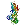

 PDBj
PDBj


 Assembly
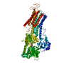
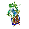
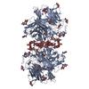
 Chlorocebus sabaeus (green monkey) / References: UniProt: P16615, P-type Ca2+ transporter


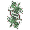
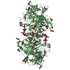
 Mass: 507.181 Da / Num. of mol.: 1 / Source method: obtained synthetically / Formula: C10H16N5O13P3 / Feature type: SUBJECT OF INVESTIGATION / Comment: ATP, energy-carrying molecule*YM
 Mass: 39.098 Da / Num. of mol.: 1 / Source method: obtained synthetically / Formula: K
 Mass: 787.121 Da / Num. of mol.: 4 / Source method: obtained synthetically / Formula: C44H85NO8P / Comment: DOPC, phospholipid*YM
 Mass: 118.174 Da / Num. of mol.: 1 / Source method: obtained synthetically / Formula: C6H14O2 / Comment: precipitant*YM
 Sample preparation
 Processing