 Movie
Movie Controller

[English] 日本語
 Yorodumi
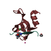
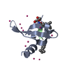
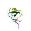
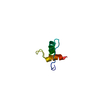
- PDB-3i91: Crystal structure of human chromobox homolog 8 (CBX8) with H3K9 p... -
+ Open data

- Basic information
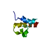
| Entry | Database: PDB / ID: 3i91 | ||||||
|---|---|---|---|---|---|---|---|
| Title | Crystal structure of human chromobox homolog 8 (CBX8) with H3K9 peptide | ||||||
 Components Components |
| ||||||
 Keywords Keywords | TRANSCRIPTION / Chromobox homolog 8 / CBX8 / H3K9 peptide / Structural Genomics / Structural Genomics Consortium / SGC / Chromatin regulator / Nucleus / Phosphoprotein / Repressor / Transcription regulation | ||||||
| Function / homology |  Function and homology information Function and homology informationPRC1 complex / histone H3K27me3 reader activity / ubiquitin-protein transferase activator activity / PcG protein complex / SUMOylation of DNA methylation proteins / SUMOylation of RNA binding proteins / positive regulation of collagen biosynthetic process / RUNX1 interacts with co-factors whose precise effect on RUNX1 targets is not known / SUMOylation of DNA damage response and repair proteins / heterochromatin ...PRC1 complex / histone H3K27me3 reader activity / ubiquitin-protein transferase activator activity / PcG protein complex / SUMOylation of DNA methylation proteins / SUMOylation of RNA binding proteins / positive regulation of collagen biosynthetic process / RUNX1 interacts with co-factors whose precise effect on RUNX1 targets is not known / SUMOylation of DNA damage response and repair proteins / heterochromatin / SUMOylation of transcription cofactors / SUMOylation of chromatin organization proteins / Regulation of PTEN gene transcription / positive regulation of DNA repair / cellular response to hydrogen peroxide / heterochromatin formation / Oxidative Stress Induced Senescence / single-stranded RNA binding / negative regulation of DNA-templated transcription / positive regulation of cell population proliferation / chromatin binding / chromatin / DNA-templated transcription / negative regulation of transcription by RNA polymerase II / nucleoplasm / nucleus Similarity search - Function | ||||||
| Biological species |  Homo sapiens (human) Homo sapiens (human) | ||||||
| Method |  X-RAY DIFFRACTION / SYNCHROTRON / MOLECULAR REPLACEMENT / Resolution: 1.55 Å X-RAY DIFFRACTION / SYNCHROTRON / MOLECULAR REPLACEMENT / Resolution: 1.55 Å | ||||||
 Authors Authors | Amaya, M.F. / Ravichandran, M. / Loppnau, P. / Kozieradzki, I. / Edwards, A.M. / Arrowsmith, C.H. / Weigelt, J. / Bountra, C. / Bochkarev, A. / Min, J. ...Amaya, M.F. / Ravichandran, M. / Loppnau, P. / Kozieradzki, I. / Edwards, A.M. / Arrowsmith, C.H. / Weigelt, J. / Bountra, C. / Bochkarev, A. / Min, J. / Ouyang, H. / Structural Genomics Consortium (SGC) | ||||||
 Citation Citation | Journal: J.Biol.Chem. / Year: 2011 Title: Recognition and specificity determinants of the human cbx chromodomains. Authors: Kaustov, L. / Ouyang, H. / Amaya, M. / Lemak, A. / Nady, N. / Duan, S. / Wasney, G.A. / Li, Z. / Vedadi, M. / Schapira, M. / Min, J. / Arrowsmith, C.H. | ||||||
| History |
|
- Structure visualization
| Structure viewer | Molecule: MolmilJmol/JSmol |
|---|
- Downloads & links
-Download
| PDBx/mmCIF format | 3i91.cif.gz | 42.1 KB | Display | PDBx/mmCIF format |
|---|---|---|---|---|
| PDB format | pdb3i91.ent.gz | 29.5 KB | Display | PDB format |
| PDBx/mmJSON format | 3i91.json.gz | Tree view | PDBx/mmJSON format | |
| Others |  Other downloads Other downloads |
-Validation report
| Arichive directory | https://data.pdbj.org/pub/pdb/validation_reports/i9/3i91ftp://data.pdbj.org/pub/pdb/validation_reports/i9/3i91 | HTTPS FTP |
|---|
-Related structure data
| Related structure data | 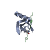 2l11C  2l12C  2l1bC 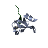 3fdtC 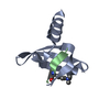 3gv6C  3h91C  3i90C  3i8y C: citing same article ( |
|---|---|
| Similar structure data |


-Links
 PDBj
PDBj


- Assembly
| Deposited unit | 
| ||||||||
|---|---|---|---|---|---|---|---|---|---|
| 1 | 
| ||||||||
| 2 | 
| ||||||||
| Unit cell |
|
-Components
| #1: Protein | Mass: 6610.602 Da / Num. of mol.: 2 / Fragment: Chromo domain: UNP residues 8-61 Source method: isolated from a genetically manipulated source Source: (gene. exp.) Homo sapiens (human) / Gene: CBX8, PC3, RC1 / Production host:  #2: Protein/peptide | | Mass: 892.013 Da / Num. of mol.: 1 / Source method: obtained synthetically / Details: Synthetic peptide #3: Water | ChemComp-HOH / |  Mass: 18.015 Da / Num. of mol.: 215 / Source method: isolated from a natural source / Formula: H2O Mass: 18.015 Da / Num. of mol.: 215 / Source method: isolated from a natural source / Formula: H2OHas protein modification | Y | |
|---|
-Experimental details
-Experiment
| Experiment | Method: X-RAY DIFFRACTION / Number of used crystals: 1 |
|---|
- Sample preparation
| Crystal | Density Matthews: 2.77 Å3/Da / Density % sol: 55.68 % |
|---|---|
| Crystal grow | Temperature: 298 K / Method: vapor diffusion / pH: 7 / Details: pH 7.0, VAPOR DIFFUSION, temperature 298K |
-Data collection
| Diffraction | Mean temperature: 100 K |
|---|---|
| Diffraction source | Source: SYNCHROTRON / Site: CLSI  / Beamline: 08ID-1 / Wavelength: 1 Å / Beamline: 08ID-1 / Wavelength: 1 Å |
| Detector | Type: MARMOSAIC 225 mm CCD / Detector: CCD Details: Cryo-cooled first crystal and sagitally focusing second crystal |
| Radiation | Monochromator: Si(111) double crystal / Protocol: SINGLE WAVELENGTH / Monochromatic (M) / Laue (L): M / Scattering type: x-ray |
| Radiation wavelength | Wavelength: 1 Å / Relative weight: 1 |
| Reflection | Resolution: 1.55→50 Å / Num. obs: 23019 |
- Processing
| Software |
| |||||||||||||||||||||||||||||||||||||||||||||||||||||||||||||||||
|---|---|---|---|---|---|---|---|---|---|---|---|---|---|---|---|---|---|---|---|---|---|---|---|---|---|---|---|---|---|---|---|---|---|---|---|---|---|---|---|---|---|---|---|---|---|---|---|---|---|---|---|---|---|---|---|---|---|---|---|---|---|---|---|---|---|---|
| Refinement | Method to determine structure: MOLECULAR REPLACEMENT / Resolution: 1.55→50 Å / Cor.coef. Fo:Fc: 0.945 / Cor.coef. Fo:Fc free: 0.927 / Occupancy max: 1 / Occupancy min: 1 / SU B: 1.415 / SU ML: 0.053 / Cross valid method: THROUGHOUT / σ(F): 0 / ESU R: 0.088 / ESU R Free: 0.091 / Stereochemistry target values: MAXIMUM LIKELIHOOD Details: 1. HYDROGENS HAVE BEEN ADDED IN THE RIDING POSITIONS. 2. U VALUES: REFINED INDIVIDUALLY.
| |||||||||||||||||||||||||||||||||||||||||||||||||||||||||||||||||
| Solvent computation | Ion probe radii: 0.8 Å / Shrinkage radii: 0.8 Å / VDW probe radii: 1.4 Å / Solvent model: MASK | |||||||||||||||||||||||||||||||||||||||||||||||||||||||||||||||||
| Displacement parameters | Biso max: 67.95 Å2 / Biso mean: 19.565 Å2 / Biso min: 6.53 Å2
| |||||||||||||||||||||||||||||||||||||||||||||||||||||||||||||||||
| Refinement step | Cycle: LAST / Resolution: 1.55→50 Å
| |||||||||||||||||||||||||||||||||||||||||||||||||||||||||||||||||
| Refine LS restraints |
| |||||||||||||||||||||||||||||||||||||||||||||||||||||||||||||||||
| LS refinement shell | Resolution: 1.55→1.59 Å / Total num. of bins used: 20
|
