 Movie
Movie Controller

[English] 日本語
 Yorodumi
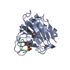
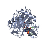
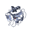
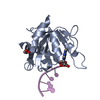
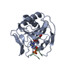
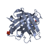
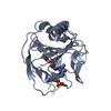
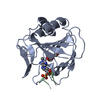
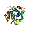
- PDB-3i3q: Crystal Structure of AlkB in complex with Mn(II) and 2-oxoglutarate -
+ Open data

- Basic information
| Entry | Database: PDB / ID: 3i3q | ||||||
|---|---|---|---|---|---|---|---|
| Title | Crystal Structure of AlkB in complex with Mn(II) and 2-oxoglutarate | ||||||
 Components Components | Alpha-ketoglutarate-dependent dioxygenase alkB | ||||||
 Keywords Keywords | OXIDOREDUCTASE / beta jellyroll / Dioxygenase / DNA damage / DNA repair / Iron / Metal-binding | ||||||
| Function / homology |  Function and homology information Function and homology informationresponse to methyl methanesulfonate / oxidative RNA demethylation / DNA oxidative demethylase / broad specificity oxidative DNA demethylase activity / oxidative RNA demethylase activity / RNA repair / oxidative demethylation / DNA alkylation repair / dioxygenase activity / ferrous iron binding ...response to methyl methanesulfonate / oxidative RNA demethylation / DNA oxidative demethylase / broad specificity oxidative DNA demethylase activity / oxidative RNA demethylase activity / RNA repair / oxidative demethylation / DNA alkylation repair / dioxygenase activity / ferrous iron binding / DNA repair / cytoplasm / cytosol Similarity search - Function | ||||||
| Biological species |  | ||||||
| Method |  X-RAY DIFFRACTION / SYNCHROTRON / MOLECULAR REPLACEMENT / molecular replacement / Resolution: 1.4 Å X-RAY DIFFRACTION / SYNCHROTRON / MOLECULAR REPLACEMENT / molecular replacement / Resolution: 1.4 Å | ||||||
 Authors Authors | Yu, B. / Hunt, J.F. | ||||||
 Citation Citation | Journal: Proc.Natl.Acad.Sci.USA / Year: 2009 Title: Enzymological and structural studies of the mechanism of promiscuous substrate recognition by the oxidative DNA repair enzyme AlkB. Authors: Yu, B. / Hunt, J.F. | ||||||
| History |
|
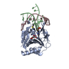
- Structure visualization
| Structure viewer | Molecule: MolmilJmol/JSmol |
|---|
- Downloads & links
-Download
| PDBx/mmCIF format | 3i3q.cif.gz | 100.2 KB | Display | PDBx/mmCIF format |
|---|---|---|---|---|
| PDB format | pdb3i3q.ent.gz | 75.5 KB | Display | PDB format |
| PDBx/mmJSON format | 3i3q.json.gz | Tree view | PDBx/mmJSON format | |
| Others |  Other downloads Other downloads |
-Validation report
| Arichive directory | https://data.pdbj.org/pub/pdb/validation_reports/i3/3i3qftp://data.pdbj.org/pub/pdb/validation_reports/i3/3i3q | HTTPS FTP |
|---|
-Related structure data


-Links
 PDBj
PDBj

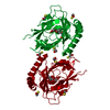
- Assembly
| Deposited unit | 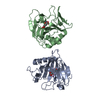
| ||||||||
|---|---|---|---|---|---|---|---|---|---|
| 1 | 
| ||||||||
| 2 | 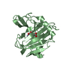
| ||||||||
| Unit cell |
|
-Components
| #1: Protein | Mass: 23610.975 Da / Num. of mol.: 2 / Fragment: UNP residues 12-216 Source method: isolated from a genetically manipulated source Source: (gene. exp.) References: UniProt: P05050, Oxidoreductases; Acting on paired donors, with incorporation or reduction of molecular oxygen; With 2-oxoglutarate as one donor, and incorporation of one atom of oxygen into each donor #2: Chemical |   Mass: 146.098 Da / Num. of mol.: 2 / Source method: obtained synthetically / Formula: C5H6O5 Mass: 146.098 Da / Num. of mol.: 2 / Source method: obtained synthetically / Formula: C5H6O5#3: Chemical |   Mass: 54.938 Da / Num. of mol.: 2 / Source method: obtained synthetically / Formula: Mn Mass: 54.938 Da / Num. of mol.: 2 / Source method: obtained synthetically / Formula: Mn#4: Water | ChemComp-HOH / |  Mass: 18.015 Da / Num. of mol.: 336 / Source method: isolated from a natural source / Formula: H2O Mass: 18.015 Da / Num. of mol.: 336 / Source method: isolated from a natural source / Formula: H2O |
|---|
-Experimental details
-Experiment
| Experiment | Method: X-RAY DIFFRACTION / Number of used crystals: 1 |
|---|
- Sample preparation
| Crystal | Density Matthews: 2.12 Å3/Da / Density % sol: 42 % |
|---|---|
| Crystal grow | Temperature: 298 K / Method: vapor diffusion, hanging drop Details: PEG 3350, sodium formate, VAPOR DIFFUSION, HANGING DROP, temperature 298K |
-Data collection
| Diffraction source | Source: SYNCHROTRON / Site: NSLS  / Beamline: X4C / Wavelength: 0.97947 Å / Beamline: X4C / Wavelength: 0.97947 Å | |||||||||||||||||||||||||||||||||||||||||||||||||||||||||||||||||||||||||||||||||||||||||||||||||||||||||
|---|---|---|---|---|---|---|---|---|---|---|---|---|---|---|---|---|---|---|---|---|---|---|---|---|---|---|---|---|---|---|---|---|---|---|---|---|---|---|---|---|---|---|---|---|---|---|---|---|---|---|---|---|---|---|---|---|---|---|---|---|---|---|---|---|---|---|---|---|---|---|---|---|---|---|---|---|---|---|---|---|---|---|---|---|---|---|---|---|---|---|---|---|---|---|---|---|---|---|---|---|---|---|---|---|---|---|
| Detector | Detector: CCD / Date: Nov 15, 2006 | |||||||||||||||||||||||||||||||||||||||||||||||||||||||||||||||||||||||||||||||||||||||||||||||||||||||||
| Radiation | Protocol: SINGLE WAVELENGTH / Scattering type: x-ray | |||||||||||||||||||||||||||||||||||||||||||||||||||||||||||||||||||||||||||||||||||||||||||||||||||||||||
| Radiation wavelength | Wavelength: 0.97947 Å / Relative weight: 1 | |||||||||||||||||||||||||||||||||||||||||||||||||||||||||||||||||||||||||||||||||||||||||||||||||||||||||
| Reflection | Resolution: 1.4→50 Å / Num. obs: 71196 / % possible obs: 96.1 % / Redundancy: 4 % / Biso Wilson estimate: 16.3 Å2 / Rmerge(I) obs: 0.047 / Net I/σ(I): 11.8 | |||||||||||||||||||||||||||||||||||||||||||||||||||||||||||||||||||||||||||||||||||||||||||||||||||||||||
| Reflection shell |
|
-Phasing
| Phasing | Method: molecular replacement | ||||||
|---|---|---|---|---|---|---|---|
| Phasing MR | Rfactor: 45.3 / Cor.coef. Fo:Fc: 40.13
|
- Processing
| Software |
| ||||||||||||||||||||||||||||||||||||||||||||||||||||||||||||||||||||||||||||||||
|---|---|---|---|---|---|---|---|---|---|---|---|---|---|---|---|---|---|---|---|---|---|---|---|---|---|---|---|---|---|---|---|---|---|---|---|---|---|---|---|---|---|---|---|---|---|---|---|---|---|---|---|---|---|---|---|---|---|---|---|---|---|---|---|---|---|---|---|---|---|---|---|---|---|---|---|---|---|---|---|---|---|
| Refinement | Method to determine structure: MOLECULAR REPLACEMENT / Resolution: 1.4→32.97 Å / Rfactor Rfree error: 0.003 / Occupancy max: 1 / Occupancy min: 0.2 / Data cutoff high absF: 792490.79 / Data cutoff low absF: 0 / Isotropic thermal model: RESTRAINED / Cross valid method: THROUGHOUT / σ(F): 0 / Details: BULK SOLVENT MODEL USED
| ||||||||||||||||||||||||||||||||||||||||||||||||||||||||||||||||||||||||||||||||
| Solvent computation | Solvent model: FLAT MODEL / Bsol: 50.8179 Å2 / ksol: 0.4 e/Å3 | ||||||||||||||||||||||||||||||||||||||||||||||||||||||||||||||||||||||||||||||||
| Displacement parameters | Biso mean: 17.5 Å2
| ||||||||||||||||||||||||||||||||||||||||||||||||||||||||||||||||||||||||||||||||
| Refine analyze |
| ||||||||||||||||||||||||||||||||||||||||||||||||||||||||||||||||||||||||||||||||
| Refinement step | Cycle: LAST / Resolution: 1.4→32.97 Å
| ||||||||||||||||||||||||||||||||||||||||||||||||||||||||||||||||||||||||||||||||
| Refine LS restraints |
| ||||||||||||||||||||||||||||||||||||||||||||||||||||||||||||||||||||||||||||||||
| Refine LS restraints NCS | NCS model details: NONE | ||||||||||||||||||||||||||||||||||||||||||||||||||||||||||||||||||||||||||||||||
| LS refinement shell | Resolution: 1.4→1.49 Å / Rfactor Rfree error: 0.008 / Total num. of bins used: 6
| ||||||||||||||||||||||||||||||||||||||||||||||||||||||||||||||||||||||||||||||||
| Xplor file |
|
