 Movie
Movie Controller

[English] 日本語
 Yorodumi
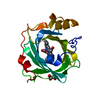
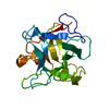
- PDB-4xmg: Crystal structure of nitrophorin 7 from Rhodnius prolixus at pH 7... -
+ Open data

- Basic information
| Entry | Database: PDB / ID: 4xmg | ||||||
|---|---|---|---|---|---|---|---|
| Title | Crystal structure of nitrophorin 7 from Rhodnius prolixus at pH 7.8 complexed with imidazole | ||||||
 Components Components | Nitrophorin-7 | ||||||
 Keywords Keywords | TRANSPORT PROTEIN / Heme / NO transporter / oxidoreductase | ||||||
| Function / homology |  Function and homology information Function and homology informationvenom-mediated perturbation of biological process / modulation of process of another organism / nitrite dismutase / histamine binding / nitric oxide binding / vasodilation / toxin activity / oxidoreductase activity / extracellular region / metal ion binding Similarity search - Function | ||||||
| Biological species |  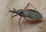 Rhodnius prolixus (insect) Rhodnius prolixus (insect) | ||||||
| Method |  X-RAY DIFFRACTION / SYNCHROTRON / MOLECULAR REPLACEMENT / Resolution: 1.8 Å X-RAY DIFFRACTION / SYNCHROTRON / MOLECULAR REPLACEMENT / Resolution: 1.8 Å | ||||||
 Authors Authors | Ogata, H. | ||||||
 Citation Citation | Journal: F1000Res / Year: 2015 Title: Structure and dynamics of the membrane attaching nitric oxide transporter nitrophorin 7. Authors: Knipp, M. / Ogata, H. / Soavi, G. / Cerullo, G. / Allegri, A. / Abbruzzetti, S. / Bruno, S. / Viappiani, C. / Bidon-Chanal, A. / Luque, F.J. | ||||||
| History |
|
- Structure visualization
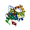
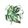
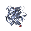
| Structure viewer | Molecule: MolmilJmol/JSmol |
|---|
- Downloads & links
-Download
| PDBx/mmCIF format | 4xmg.cif.gz | 55.9 KB | Display | PDBx/mmCIF format |
|---|---|---|---|---|
| PDB format | pdb4xmg.ent.gz | 38.1 KB | Display | PDB format |
| PDBx/mmJSON format | 4xmg.json.gz | Tree view | PDBx/mmJSON format | |
| Others |  Other downloads Other downloads |
-Validation report
| Arichive directory | https://data.pdbj.org/pub/pdb/validation_reports/xm/4xmgftp://data.pdbj.org/pub/pdb/validation_reports/xm/4xmg | HTTPS FTP |
|---|
-Related structure data
| Related structure data |  4xmcC 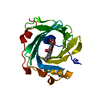 4xmdC  4xmeC  4xmfC 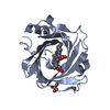 4xmhC 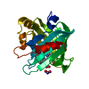 3mvfS S: Starting model for refinement C: citing same article ( |
|---|---|
| Similar structure data |


-Links
 PDBj
PDBj


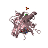
- Assembly
| Deposited unit | 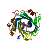
| ||||||||
|---|---|---|---|---|---|---|---|---|---|
| 1 |
| ||||||||
| Unit cell |
|
-Components
| #1: Protein | Mass: 20759.660 Da / Num. of mol.: 1 / Fragment: UNP residues 22-105 Source method: isolated from a genetically manipulated source Source: (gene. exp.) Rhodnius prolixus (insect) / Production host:  |
|---|---|
| #2: Chemical | ChemComp-HEM /   Mass: 616.487 Da / Num. of mol.: 1 / Source method: obtained synthetically / Formula: C34H32FeN4O4 Mass: 616.487 Da / Num. of mol.: 1 / Source method: obtained synthetically / Formula: C34H32FeN4O4 |
| #3: Chemical | ChemComp-IMD /   Mass: 69.085 Da / Num. of mol.: 1 / Source method: obtained synthetically / Formula: C3H5N2 Mass: 69.085 Da / Num. of mol.: 1 / Source method: obtained synthetically / Formula: C3H5N2 |
| #4: Water | ChemComp-HOH /  Mass: 18.015 Da / Num. of mol.: 120 / Source method: isolated from a natural source / Formula: H2O Mass: 18.015 Da / Num. of mol.: 120 / Source method: isolated from a natural source / Formula: H2O |
| Has protein modification | Y |
-Experimental details
-Experiment
| Experiment | Method: X-RAY DIFFRACTION |
|---|
- Sample preparation
| Crystal | Density Matthews: 2.12 Å3/Da / Density % sol: 42.06 % |
|---|---|
| Crystal grow | Temperature: 277 K / Method: vapor diffusion, sitting drop Details: 25%(w/v) PEG 3350, 0.1 M bis-Tris propane pH 7.8. 0.25%(w/v) hexamminecobalt(III) chloride, 0.25%(w/v) salicylamide, 0.25%(w/v) sulfanilamide, 0.25%(w/v) vanilic acid, 0.02 M HEPES sodium pH 6.8. |
-Data collection
| Diffraction | Mean temperature: 100 K |
|---|---|
| Diffraction source | Source: SYNCHROTRON / Site: SLS  / Beamline: X06SA / Wavelength: 0.8 Å / Beamline: X06SA / Wavelength: 0.8 Å |
| Detector | Type: PSI PILATUS 6M / Detector: PIXEL / Date: Dec 20, 2012 |
| Radiation | Protocol: SINGLE WAVELENGTH / Monochromatic (M) / Laue (L): M / Scattering type: x-ray |
| Radiation wavelength | Wavelength: 0.8 Å / Relative weight: 1 |
| Reflection | Resolution: 1.8→34.25 Å / Num. obs: 15836 / % possible obs: 97.6 % / Redundancy: 3.3 % / Rmerge(I) obs: 0.083 / Net I/σ(I): 9.2 |
| Reflection shell | Resolution: 1.8→1.85 Å / Rmerge(I) obs: 0.439 / Mean I/σ(I) obs: 3 / % possible all: 96.9 |
- Processing
| Software |
| |||||||||||||||||||||||||||||||||||||||||||||||||
|---|---|---|---|---|---|---|---|---|---|---|---|---|---|---|---|---|---|---|---|---|---|---|---|---|---|---|---|---|---|---|---|---|---|---|---|---|---|---|---|---|---|---|---|---|---|---|---|---|---|---|
| Refinement | Method to determine structure: MOLECULAR REPLACEMENT Starting model: 3MVF Resolution: 1.8→34.245 Å / SU ML: 0.43 / Cross valid method: FREE R-VALUE / σ(F): 2 / Phase error: 22.62 / Stereochemistry target values: ML
| |||||||||||||||||||||||||||||||||||||||||||||||||
| Solvent computation | Shrinkage radii: 0.86 Å / VDW probe radii: 1.1 Å / Solvent model: FLAT BULK SOLVENT MODEL / Bsol: 36.634 Å2 / ksol: 0.338 e/Å3 | |||||||||||||||||||||||||||||||||||||||||||||||||
| Displacement parameters |
| |||||||||||||||||||||||||||||||||||||||||||||||||
| Refinement step | Cycle: LAST / Resolution: 1.8→34.245 Å
| |||||||||||||||||||||||||||||||||||||||||||||||||
| Refine LS restraints |
| |||||||||||||||||||||||||||||||||||||||||||||||||
| LS refinement shell |
|
