 Movie
Movie Controller


+ Open data

- Basic information
| Entry | Database: PDB / ID: 3i3f | ||||||
|---|---|---|---|---|---|---|---|
| Title | Hypothetical protein from Giardia lamblia GL50803_14299 | ||||||
 Components Components | Hypothetical protein | ||||||
 Keywords Keywords | UNKNOWN FUNCTION / hypothetical / structural genomics / NIAID / deCODE / infectious disease / Seattle Structural Genomics Center for Infectious Disease / SSGCID | ||||||
| Function / homology |  Function and homology information Function and homology information | ||||||
| Biological species |  Giardia lamblia (eukaryote) Giardia lamblia (eukaryote) | ||||||
| Method |  X-RAY DIFFRACTION / SYNCHROTRON / MOLECULAR REPLACEMENT / molecular replacement / Resolution: 1.35 Å X-RAY DIFFRACTION / SYNCHROTRON / MOLECULAR REPLACEMENT / molecular replacement / Resolution: 1.35 Å | ||||||
 Authors Authors | Seattle Structural Genomics Center for Infectious Disease (SSGCID) | ||||||
 Citation Citation | Journal: To be Published Title: Hypothetical protein from Giardia lamblia GL50803_14299 Authors: Arakaki, T.L. / Abendroth, J. / Staker, B.L. | ||||||
| History |
|
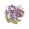
- Structure visualization
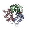
| Structure viewer | Molecule: MolmilJmol/JSmol |
|---|
- Downloads & links
-Download
| PDBx/mmCIF format | 3i3f.cif.gz | 97 KB | Display | PDBx/mmCIF format |
|---|---|---|---|---|
| PDB format | pdb3i3f.ent.gz | 74.2 KB | Display | PDB format |
| PDBx/mmJSON format | 3i3f.json.gz | Tree view | PDBx/mmJSON format | |
| Others |  Other downloads Other downloads |
-Validation report
| Arichive directory | https://data.pdbj.org/pub/pdb/validation_reports/i3/3i3fftp://data.pdbj.org/pub/pdb/validation_reports/i3/3i3f | HTTPS FTP |
|---|
-Related structure data
| Similar structure data | |
|---|---|
| Other databases |


-Links
 PDBj
PDBj
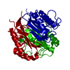
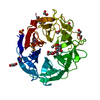
- Assembly
| Deposited unit | 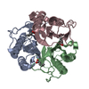
| ||||||||
|---|---|---|---|---|---|---|---|---|---|
| 1 |
| ||||||||
| Unit cell |
| ||||||||
| Components on special symmetry positions |
|
-Components
| #1: Protein | Mass: 15567.940 Da / Num. of mol.: 3 Source method: isolated from a genetically manipulated source Source: (gene. exp.) Giardia lamblia (eukaryote) / Strain: ATCC 50803 / Gene: GL50803_14299 / Plasmid: AVA0421 / Production host:  #2: Chemical | 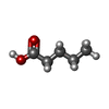  Mass: 102.132 Da / Num. of mol.: 2 / Source method: obtained synthetically / Formula: C5H10O2 Mass: 102.132 Da / Num. of mol.: 2 / Source method: obtained synthetically / Formula: C5H10O2#3: Chemical | ChemComp-BUA / |   Mass: 88.105 Da / Num. of mol.: 1 / Source method: obtained synthetically / Formula: C4H8O2 Mass: 88.105 Da / Num. of mol.: 1 / Source method: obtained synthetically / Formula: C4H8O2#4: Water | ChemComp-HOH / |  Mass: 18.015 Da / Num. of mol.: 443 / Source method: isolated from a natural source / Formula: H2O Mass: 18.015 Da / Num. of mol.: 443 / Source method: isolated from a natural source / Formula: H2OHas protein modification | Y | Nonpolymer details | AUTHOR STATE THA THESE GROUPS WERE MODELED IN RESIDUAL ELECTRON DENSITY BASED UPON THE BEST BIT, ...AUTHOR STATE THA THESE GROUPS WERE MODELED IN RESIDUAL ELECTRON DENSITY BASED UPON THE BEST BIT, BUT THE EXACT IDENTITY OF THE LIGANDS MAY NOT BE CORRECT. | |
|---|
-Experimental details
-Experiment
| Experiment | Method: X-RAY DIFFRACTION / Number of used crystals: 1 |
|---|
- Sample preparation
| Crystal | Density Matthews: 2.17 Å3/Da / Density % sol: 38.87 % |
|---|---|
| Crystal grow | Temperature: 290 K / Method: sitting drop, vapor diffusion / pH: 5.5 Details: 100 mM Tris, pH 5.5, 25% PEG 3350, 200 mM ammonium acetate, sitting drop, vapor diffusion, temperature 290K |
-Data collection
| Diffraction | Mean temperature: 100 K | ||||||||||||||||||||||||||||||||||||||||||||||||||||||||||||||||||||||||||||||||||||||||
|---|---|---|---|---|---|---|---|---|---|---|---|---|---|---|---|---|---|---|---|---|---|---|---|---|---|---|---|---|---|---|---|---|---|---|---|---|---|---|---|---|---|---|---|---|---|---|---|---|---|---|---|---|---|---|---|---|---|---|---|---|---|---|---|---|---|---|---|---|---|---|---|---|---|---|---|---|---|---|---|---|---|---|---|---|---|---|---|---|---|
| Diffraction source | Source: SYNCHROTRON / Site: SSRL  / Beamline: BL12-2 / Wavelength: 0.9795 Å / Beamline: BL12-2 / Wavelength: 0.9795 Å | ||||||||||||||||||||||||||||||||||||||||||||||||||||||||||||||||||||||||||||||||||||||||
| Detector | Type: ADSC Quantum-315R CCD / Detector: CCD / Date: Jan 1, 2009 | ||||||||||||||||||||||||||||||||||||||||||||||||||||||||||||||||||||||||||||||||||||||||
| Radiation | Protocol: SINGLE WAVELENGTH / Monochromatic (M) / Laue (L): M / Scattering type: x-ray | ||||||||||||||||||||||||||||||||||||||||||||||||||||||||||||||||||||||||||||||||||||||||
| Radiation wavelength | Wavelength: 0.9795 Å / Relative weight: 1 | ||||||||||||||||||||||||||||||||||||||||||||||||||||||||||||||||||||||||||||||||||||||||
| Reflection | Resolution: 1.35→39.41 Å / Num. all: 82643 / Num. obs: 82643 / % possible obs: 99.5 % / Redundancy: 6.3 % / Rmerge(I) obs: 0.074 / Rsym value: 0.074 / Net I/σ(I): 13.5 | ||||||||||||||||||||||||||||||||||||||||||||||||||||||||||||||||||||||||||||||||||||||||
| Reflection shell | Diffraction-ID: 1
|
-Phasing
| Phasing | Method: molecular replacement | |||||||||
|---|---|---|---|---|---|---|---|---|---|---|
| Phasing MR |
|
- Processing
| Software |
| |||||||||||||||||||||||||||||||||||||||||||||||||||||||||||||||||||||||||||||||||||||
|---|---|---|---|---|---|---|---|---|---|---|---|---|---|---|---|---|---|---|---|---|---|---|---|---|---|---|---|---|---|---|---|---|---|---|---|---|---|---|---|---|---|---|---|---|---|---|---|---|---|---|---|---|---|---|---|---|---|---|---|---|---|---|---|---|---|---|---|---|---|---|---|---|---|---|---|---|---|---|---|---|---|---|---|---|---|---|
| Refinement | Method to determine structure: MOLECULAR REPLACEMENT / Resolution: 1.35→37.35 Å / Cor.coef. Fo:Fc: 0.967 / Cor.coef. Fo:Fc free: 0.959 / WRfactor Rfree: 0.201 / WRfactor Rwork: 0.187 / Occupancy max: 1 / Occupancy min: 0.25 / FOM work R set: 0.89 / SU B: 0.872 / SU ML: 0.036 / SU R Cruickshank DPI: 0.056 / SU Rfree: 0.056 / Cross valid method: THROUGHOUT / σ(F): 0 / ESU R: 0.056 / ESU R Free: 0.056 / Stereochemistry target values: MAXIMUM LIKELIHOOD Details: HYDROGENS HAVE BEEN ADDED IN THE RIDING POSITIONS U VALUES : REFINED INDIVIDUALLY
| |||||||||||||||||||||||||||||||||||||||||||||||||||||||||||||||||||||||||||||||||||||
| Solvent computation | Ion probe radii: 0.8 Å / Shrinkage radii: 0.8 Å / VDW probe radii: 1.4 Å / Solvent model: BABINET MODEL WITH MASK | |||||||||||||||||||||||||||||||||||||||||||||||||||||||||||||||||||||||||||||||||||||
| Displacement parameters | Biso max: 56.86 Å2 / Biso mean: 14.501 Å2 / Biso min: 7.77 Å2
| |||||||||||||||||||||||||||||||||||||||||||||||||||||||||||||||||||||||||||||||||||||
| Refinement step | Cycle: LAST / Resolution: 1.35→37.35 Å
| |||||||||||||||||||||||||||||||||||||||||||||||||||||||||||||||||||||||||||||||||||||
| Refine LS restraints |
| |||||||||||||||||||||||||||||||||||||||||||||||||||||||||||||||||||||||||||||||||||||
| LS refinement shell | Resolution: 1.35→1.385 Å / Total num. of bins used: 20
|
