 Movie
Movie Controller


+ Open data

- Basic information
| Entry | Database: PDB / ID: 1jd1 | ||||||
|---|---|---|---|---|---|---|---|
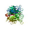
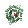
| Title | Crystal Structure of YEO7_yeast | ||||||
 Components Components | HYPOTHETICAL 13.9 KDA PROTEIN IN FCY2-PET117 INTERGENIC REGION | ||||||
 Keywords Keywords | TRANSLATION INHIBITOR / STRUCTURAL GENOMICS / PSI / Protein Structure Initiative / New York SGX Research Center for Structural Genomics / NYSGXRC | ||||||
| Function / homology |  Function and homology information Function and homology informationdeaminase activity / mitochondrial intermembrane space / mitochondrion / nucleus / cytosol / cytoplasm Similarity search - Function | ||||||
| Biological species |  | ||||||
| Method |  X-RAY DIFFRACTION / SYNCHROTRON / MAD / Resolution: 1.7 Å X-RAY DIFFRACTION / SYNCHROTRON / MAD / Resolution: 1.7 Å | ||||||
 Authors Authors | Deaconescu, A.M. / Burley, S.K. / New York SGX Research Center for Structural Genomics (NYSGXRC) | ||||||
 Citation Citation | Journal: Proteins / Year: 2002 Title: X-ray structure of Saccharomyces cerevisiae homologous mitochondrial matrix factor 1 (Hmf1). Authors: Deaconescu, A.M. / Roll-Mecak, A. / Bonanno, J.B. / Gerchman, S.E. / Kycia, H. / Studier, F.W. / Burley, S.K. | ||||||
| History |
|
- Structure visualization
| Structure viewer | Molecule: MolmilJmol/JSmol |
|---|
- Downloads & links
-Download
| PDBx/mmCIF format | 1jd1.cif.gz | 155.9 KB | Display | PDBx/mmCIF format |
|---|---|---|---|---|
| PDB format | pdb1jd1.ent.gz | 124.3 KB | Display | PDB format |
| PDBx/mmJSON format | 1jd1.json.gz | Tree view | PDBx/mmJSON format | |
| Others |  Other downloads Other downloads |
-Validation report
| Arichive directory | https://data.pdbj.org/pub/pdb/validation_reports/jd/1jd1ftp://data.pdbj.org/pub/pdb/validation_reports/jd/1jd1 | HTTPS FTP |
|---|
-Related structure data
| Similar structure data | |
|---|---|
| Other databases |


-Links
 PDBj
PDBj
- Assembly
| Deposited unit | 
| ||||||||
|---|---|---|---|---|---|---|---|---|---|
| 1 | 
| ||||||||
| 2 | 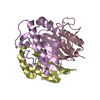
| ||||||||
| Unit cell |
| ||||||||
| Details | The biological assembly is a trimer. |
-Components
| #1: Protein | Mass: 13918.897 Da / Num. of mol.: 6 Source method: isolated from a genetically manipulated source Source: (gene. exp.) Gene: YERO57C / Species (production host): Escherichia coli / Production host:  #2: Water | ChemComp-HOH / |  Mass: 18.015 Da / Num. of mol.: 634 / Source method: isolated from a natural source / Formula: H2O Mass: 18.015 Da / Num. of mol.: 634 / Source method: isolated from a natural source / Formula: H2O |
|---|
-Experimental details
-Experiment
| Experiment | Method: X-RAY DIFFRACTION / Number of used crystals: 1 |
|---|
- Sample preparation
| Crystal | Density Matthews: 2.42 Å3/Da / Density % sol: 49.27 % | ||||||||||||||||||||||||||||||||||||||||||
|---|---|---|---|---|---|---|---|---|---|---|---|---|---|---|---|---|---|---|---|---|---|---|---|---|---|---|---|---|---|---|---|---|---|---|---|---|---|---|---|---|---|---|---|
| Crystal grow | *PLUS Temperature: 20 ℃ / pH: 7.5 / Method: vapor diffusion, hanging drop | ||||||||||||||||||||||||||||||||||||||||||
| Components of the solutions | *PLUS
|
-Data collection
| Diffraction | Mean temperature: 170 K | |||||||||
|---|---|---|---|---|---|---|---|---|---|---|
| Diffraction source | Source: SYNCHROTRON / Site: NSLS  / Beamline: X9B / Wavelength: 0.98091, 0.99104 / Beamline: X9B / Wavelength: 0.98091, 0.99104 | |||||||||
| Detector | Type: MARRESEARCH / Detector: CCD / Date: Aug 24, 2000 | |||||||||
| Radiation | Protocol: MAD / Monochromatic (M) / Laue (L): M / Scattering type: x-ray | |||||||||
| Radiation wavelength |
| |||||||||
| Reflection | Resolution: 1.7→50 Å / Num. obs: 86368 / % possible obs: 99 % / Observed criterion σ(F): 0 / Redundancy: 4 % / Biso Wilson estimate: 20.16 Å2 / Rsym value: 0.035 / Net I/σ(I): 30 | |||||||||
| Reflection shell | Resolution: 1.7→1.74 Å / Redundancy: 4 % / Mean I/σ(I) obs: 4.4 / Rsym value: 0.307 / % possible all: 97.7 | |||||||||
| Reflection | *PLUS Num. obs: 88185 / % possible obs: 98.7 % / Num. measured all: 1452497 / Rmerge(I) obs: 0.039 | |||||||||
| Reflection shell | *PLUS % possible obs: 97.1 % / Rmerge(I) obs: 0.333 |
- Processing
| Software |
| ||||||||||||||||||||
|---|---|---|---|---|---|---|---|---|---|---|---|---|---|---|---|---|---|---|---|---|---|
| Refinement | Method to determine structure: MAD / Resolution: 1.7→50 Å / Isotropic thermal model: anisotropic / σ(F): 2 / Stereochemistry target values: Engh & Huber
| ||||||||||||||||||||
| Refinement step | Cycle: LAST / Resolution: 1.7→50 Å
| ||||||||||||||||||||
| Refine LS restraints |
| ||||||||||||||||||||
| LS refinement shell | Resolution: 1.7→1.71 Å
| ||||||||||||||||||||
| Software | *PLUS Name: CNS / Classification: refinement | ||||||||||||||||||||
| Refinement | *PLUS Highest resolution: 1.7 Å / Lowest resolution: 50 Å / σ(F): 2 / Rfactor obs: 0.207 | ||||||||||||||||||||
| Solvent computation | *PLUS | ||||||||||||||||||||
| Displacement parameters | *PLUS | ||||||||||||||||||||
| Refine LS restraints | *PLUS Type: c_angle_deg / Dev ideal: 1.4 | ||||||||||||||||||||
| LS refinement shell | *PLUS Rfactor Rfree: 0.271 / Rfactor Rwork: 0.241 |
