 Movie
Movie Controller

[English] 日本語
 Yorodumi
- PDB-3a71: High resolution structure of Penicillium chrysogenum alpha-L-arab... -
+ Open data

- Basic information
| Entry | Database: PDB / ID: 3a71 | ||||||
|---|---|---|---|---|---|---|---|
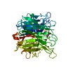
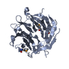
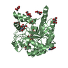
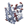
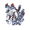
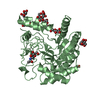
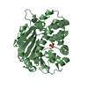
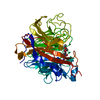
| Title | High resolution structure of Penicillium chrysogenum alpha-L-arabinanase | ||||||
 Components Components | Exo-arabinanase | ||||||
 Keywords Keywords | HYDROLASE / ARABINASE / GLYCOSYL HYDROLASE | ||||||
| Function / homology | Neuraminidase - #10 / 6 Propeller / Neuraminidase / Mainly Beta / ACETATE ION / Exo-arabinanase Function and homology information Function and homology information | ||||||
| Biological species |  Penicillium chrysogenum (fungus) Penicillium chrysogenum (fungus) | ||||||
| Method |  X-RAY DIFFRACTION / SYNCHROTRON / MAD / Resolution: 1.14 Å X-RAY DIFFRACTION / SYNCHROTRON / MAD / Resolution: 1.14 Å | ||||||
 Authors Authors | Sogabe, Y. | ||||||
 Citation Citation | Journal: Acta Crystallogr.,Sect.D / Year: 2011 Title: High-resolution structure of exo-arabinanase from Penicillium chrysogenum Authors: Sogabe, Y. / Kitatani, T. / Yamaguchi, A. / Kinoshita, T. / Adachi, H. / Takano, K. / Inoue, T. / Mori, Y. / Matsumura, H. / Sakamoto, T. / Tada, T. | ||||||
| History |
|
- Structure visualization
| Structure viewer | Molecule: MolmilJmol/JSmol |
|---|
- Downloads & links
-Download
| PDBx/mmCIF format | 3a71.cif.gz | 170.5 KB | Display | PDBx/mmCIF format |
|---|---|---|---|---|
| PDB format | pdb3a71.ent.gz | 133.7 KB | Display | PDB format |
| PDBx/mmJSON format | 3a71.json.gz | Tree view | PDBx/mmJSON format | |
| Others |  Other downloads Other downloads |
-Validation report
| Arichive directory | https://data.pdbj.org/pub/pdb/validation_reports/a7/3a71ftp://data.pdbj.org/pub/pdb/validation_reports/a7/3a71 | HTTPS FTP |
|---|
-Related structure data


-Links
 PDBj
PDBj- Assembly
| Deposited unit | 
| ||||||||
|---|---|---|---|---|---|---|---|---|---|
| 1 |
| ||||||||
| Unit cell |
|
-Components
| #1: Protein | Mass: 39359.625 Da / Num. of mol.: 1 / Fragment: UNP Residues 24-378 Source method: isolated from a genetically manipulated source Source: (gene. exp.) Penicillium chrysogenum (fungus) / Strain: 31B / Plasmid: pET3A / Production host:  References: UniProt: Q7ZA77, non-reducing end alpha-L-arabinofuranosidase | ||
|---|---|---|---|
| #2: Chemical | ChemComp-ACT /   Mass: 59.044 Da / Num. of mol.: 1 / Source method: obtained synthetically / Formula: C2H3O2 Mass: 59.044 Da / Num. of mol.: 1 / Source method: obtained synthetically / Formula: C2H3O2 | ||
| #3: Chemical |   Mass: 118.174 Da / Num. of mol.: 3 / Source method: obtained synthetically / Formula: C6H14O2 / Comment: precipitant*YM Mass: 118.174 Da / Num. of mol.: 3 / Source method: obtained synthetically / Formula: C6H14O2 / Comment: precipitant*YM#4: Water | ChemComp-HOH / |  Mass: 18.015 Da / Num. of mol.: 537 / Source method: isolated from a natural source / Formula: H2O Mass: 18.015 Da / Num. of mol.: 537 / Source method: isolated from a natural source / Formula: H2O |
-Experimental details
-Experiment
| Experiment | Method: X-RAY DIFFRACTION / Number of used crystals: 1 |
|---|
- Sample preparation
| Crystal | Density Matthews: 2.61 Å3/Da / Density % sol: 52.81 % |
|---|---|
| Crystal grow | Temperature: 277 K / pH: 4.5 Details: 28% MPD, 0.1M sodium acetate, pH 4.5, VAPOR DIFFUSION, HANGING DROP, temperature 277.0K |
-Data collection
| Diffraction | Mean temperature: 100 K | ||||||||||||
|---|---|---|---|---|---|---|---|---|---|---|---|---|---|
| Diffraction source | Source: SYNCHROTRON / Site: SPring-8  / Beamline: BL41XU / Wavelength: 0.9792, 0.9794, 0.9700 / Beamline: BL41XU / Wavelength: 0.9792, 0.9794, 0.9700 | ||||||||||||
| Detector | Type: ADSC QUANTUM 315 / Detector: CCD / Date: Nov 13, 2006 | ||||||||||||
| Radiation | Monochromator: ROTATED-INCLINED DOUBLE-CRYSTAL MO / Protocol: MAD / Monochromatic (M) / Laue (L): M / Scattering type: x-ray | ||||||||||||
| Radiation wavelength |
| ||||||||||||
| Reflection | Resolution: 1.14→10 Å / Num. obs: 149407 / % possible obs: 99.7 % / Redundancy: 7.2 % / Biso Wilson estimate: 10.7 Å2 / Rmerge(I) obs: 0.11 / Net I/σ(I): 32 | ||||||||||||
| Reflection shell | Resolution: 1.14→1.18 Å / Redundancy: 6.9 % / Rmerge(I) obs: 0.342 / Mean I/σ(I) obs: 5.4 / % possible all: 99 |
- Processing
| Software |
| |||||||||||||||||||||||||||||||||
|---|---|---|---|---|---|---|---|---|---|---|---|---|---|---|---|---|---|---|---|---|---|---|---|---|---|---|---|---|---|---|---|---|---|---|
| Refinement | Method to determine structure: MAD / Resolution: 1.14→10 Å / Cor.coef. Fo:Fc: 0.967 / Cor.coef. Fo:Fc free: 0.962 / Num. parameters: 30027 / Num. restraintsaints: 36027 / SU B: 0.338 / SU ML: 0.017 / Cross valid method: FREE R / σ(F): 0 / ESU R: 0.029 / ESU R Free: 0.03 / Stereochemistry target values: Engh & Huber / Details: ANISOTROPIC REFINEMENT REDUCED FREE R (NO CUTOFF)
| |||||||||||||||||||||||||||||||||
| Solvent computation | Ion probe radii: 0.8 Å / Shrinkage radii: 0.8 Å / VDW probe radii: 1.4 Å / Solvent model: MASK | |||||||||||||||||||||||||||||||||
| Displacement parameters | Biso mean: 8.17 Å2
| |||||||||||||||||||||||||||||||||
| Refine analyze | Num. disordered residues: 0 / Occupancy sum hydrogen: 2652 / Occupancy sum non hydrogen: 3336
| |||||||||||||||||||||||||||||||||
| Refinement step | Cycle: LAST / Resolution: 1.14→10 Å
| |||||||||||||||||||||||||||||||||
| Refine LS restraints |
| |||||||||||||||||||||||||||||||||
| LS refinement shell | Resolution: 1.14→1.17 Å / Total num. of bins used: 20
|
