[English] 日本語
Yorodumi
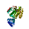
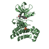
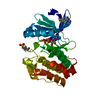
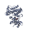
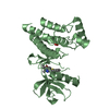
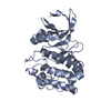
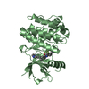
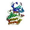
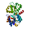
- PDB-3hx3: Crystal structure of CRALBP mutant R234W -

+
Open data


ID or keywords:

Loading...

-
Basic information

Entry
Database: PDB / ID: 3hx3
TitleCrystal structure of CRALBP mutant R234W
ComponentsRetinaldehyde-binding protein 1
KeywordsTRANSPORT PROTEIN / lipid transfer protein / 11-cis-retinal / bothnia dystrophy / Acetylation / Cytoplasm / Disease mutation / Retinitis pigmentosa / Retinol-binding / Sensory transduction / Transport / Vision
Function / homology
Function and homology information


The retinoid cycle in cones (daylight vision) / 11-cis retinal binding / vitamin A metabolic process / response to stimulus / phosphatidylinositol bisphosphate binding / retinol binding / Retinoid cycle disease events / The canonical retinoid cycle in rods (twilight vision) / visual perception / cell body ...The retinoid cycle in cones (daylight vision) / 11-cis retinal binding / vitamin A metabolic process / response to stimulus / phosphatidylinositol bisphosphate binding / retinol binding / Retinoid cycle disease events / The canonical retinoid cycle in rods (twilight vision) / visual perception / cell body / centrosome / nucleoplasm / cytosol
Similarity search - Function
N-terminal domain of phosphatidylinositol transfer protein sec14p / Phosphatidylinositol Transfer Protein Sec14p / CRAL-TRIO lipid binding domain / CRAL/TRIO, N-terminal domain / CRAL/TRIO, N-terminal domain / CRAL/TRIO, N-terminal domain / CRAL/TRIO, N-terminal domain superfamily / CRAL/TRIO domain / CRAL-TRIO lipid binding domain profile. / Domain in homologues of a S. cerevisiae phosphatidylinositol transfer protein (Sec14p) ...N-terminal domain of phosphatidylinositol transfer protein sec14p / Phosphatidylinositol Transfer Protein Sec14p / CRAL-TRIO lipid binding domain / CRAL/TRIO, N-terminal domain / CRAL/TRIO, N-terminal domain / CRAL/TRIO, N-terminal domain / CRAL/TRIO, N-terminal domain superfamily / CRAL/TRIO domain / CRAL-TRIO lipid binding domain profile. / Domain in homologues of a S. cerevisiae phosphatidylinositol transfer protein (Sec14p) / CRAL-TRIO lipid binding domain / CRAL-TRIO lipid binding domain superfamily / Helicase, Ruva Protein; domain 3 / Orthogonal Bundle / 3-Layer(aba) Sandwich / Mainly Alpha / Alpha Beta
Similarity search - Domain/homology
RETINAL / Retinaldehyde-binding protein 1
Similarity search - Component
Biological speciesHomo sapiens (human)
MethodX-RAY DIFFRACTION / SYNCHROTRON / SAD / Resolution: 1.69 Å
AuthorsStocker, A. / He, X. / Lobsiger, J.
CitationJournal: Proc.Natl.Acad.Sci.USA / Year: 2009
Title: Bothnia dystrophy is caused by domino-like rearrangements in cellular retinaldehyde-binding protein mutant R234W.
Authors: He, X. / Lobsiger, J. / Stocker, A.
History
DepositionJun 19, 2009Deposition site: RCSB / Processing site: RCSB
Revision 1.0Oct 20, 2009Provider: repository / Type: Initial release
Revision 1.1Jul 13, 2011Group: Version format compliance
Revision 1.2Oct 13, 2021Group: Database references / Derived calculations
Category: database_2 / struct_conn ...database_2 / struct_conn / struct_ref_seq_dif / struct_site
Item: _database_2.pdbx_DOI / _database_2.pdbx_database_accession ..._database_2.pdbx_DOI / _database_2.pdbx_database_accession / _struct_conn.pdbx_leaving_atom_flag / _struct_ref_seq_dif.details / _struct_site.pdbx_auth_asym_id / _struct_site.pdbx_auth_comp_id / _struct_site.pdbx_auth_seq_id

-
Structure visualization

Structure viewerMolecule:
MolmilJmol/JSmol

Downloads & links

-
Assembly

Deposited unit
A: Retinaldehyde-binding protein 1
hetero molecules


Theoretical massNumber of molelcules
Total (without water)36,9782
Polymers36,6931
Non-polymers2841
Water6,900383
1


  • Idetical with deposited unit
  • defined by author&software
TypeNameSymmetry operationNumber
identity operation1_555x,y,z1
MethodPISA
Unit cell
Length a, b, c (Å)87.930, 57.880, 75.150
Angle α, β, γ (deg.)90.000, 122.850, 90.000
Int Tables number5
Space group name H-MC121
Components on special symmetry positions
IDModelComponents
11A-401-

HOH

21A-679-

HOH

-
Components

#1: Protein Retinaldehyde-binding protein 1 / / Cellular retinaldehyde-binding protein


Mass: 36693.340 Da / Num. of mol.: 1 / Mutation: R234W
Source method: isolated from a genetically manipulated source
Source: (gene. exp.) Homo sapiens (human) / Gene: CRALBP, RLBP1 / Plasmid: pET28a / Production host: Escherichia coli (E. coli) / Strain (production host): BL21(DE3) / References: UniProt: P12271
#2: Chemical ChemComp-RET / RETINAL / Retinal


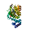
Mass: 284.436 Da / Num. of mol.: 1 / Source method: obtained synthetically / Formula: C20H28O
#3: Water ChemComp-HOH / water / Water


Mass: 18.015 Da / Num. of mol.: 383 / Source method: isolated from a natural source / Formula: H2O

-
Experimental details

-
Experiment

ExperimentMethod: X-RAY DIFFRACTION / Number of used crystals: 1

-
Sample preparation

CrystalDensity Matthews: 2.19 Å3/Da / Density % sol: 43.81 %
Crystal growTemperature: 298 K / Method: vapor diffusion / pH: 7.5
Details: PEG 3000, NaCl, pH 7.5, VAPOR DIFFUSION, temperature 298K

-
Data collection

DiffractionMean temperature: 100 K
Diffraction sourceSource: SYNCHROTRON / Site: SLS / Beamline: X06SA / Wavelength: 0.9793 Å
DetectorType: PSI PILATUS 6M / Detector: PIXEL / Date: Sep 22, 2008
RadiationMonochromator: Si(111) Channel / Protocol: SAD / Scattering type: x-ray
Radiation wavelengthWavelength: 0.9793 Å / Relative weight: 1
ReflectionResolution: 1.69→45.577 Å / Num. all: 69614 / Num. obs: 67824 / % possible obs: 97.4 % / Observed criterion σ(I): -3 / Redundancy: 5.6 % / Biso Wilson estimate: 30.96 Å2 / Rmerge(I) obs: 0.048 / Net I/σ(I): 19.8
Reflection shellResolution: 1.69→1.8 Å / Redundancy: 4.8 % / Rmerge(I) obs: 0.576 / Mean I/σ(I) obs: 2.7 / Num. unique all: 11226 / Num. unique obs: 9663 / % possible all: 86.1

-
Phasing

PhasingMethod: SAD
Phasing MADD res high: 1.78 Å / D res low: 19.45 Å / FOM : 0.53 / FOM acentric: 0.547 / FOM centric: 0.169 / Reflection: 30506 / Reflection acentric: 29134 / Reflection centric: 1362
Phasing dmFOM : 0.69 / FOM acentric: 0.7 / FOM centric: 0.52 / Reflection: 30515 / Reflection acentric: 29153 / Reflection centric: 1362
Phasing dm shell
Resolution (Å)FOM FOM acentricFOM centricReflectionReflection acentricReflection centric
5.1-19.4510.940.950.8513421187155
3.2-5.10.930.950.7340923833259
2.5-3.20.840.850.5951134867246
2.2-2.50.750.760.4451394930209
1.9-2.20.580.590.3291578840317
1.8-1.90.440.440.2856725496176

-
Processing

Software
NameVersionClassificationNB
XSCALEdata scaling
PHASERphasing
RESOLVE2.13phasing
PHENIXrefinement
PDB_EXTRACT3.005data extraction
MOSFLMdata reduction
XDSdata reduction
XDSdata scaling
RefinementMethod to determine structure: SAD / Resolution: 1.69→45.577 Å / Occupancy max: 1 / Occupancy min: 1 / SU ML: 0.2 / σ(F): 0 / Stereochemistry target values: ML
Details: The Friedel pairs were used in phasing and in refinement.
RfactorNum. reflection% reflectionSelection details
Rfree0.184 6775 9.99 %RANDOM
Rwork0.167 ---
all0.169 67814 --
obs0.169 67814 97.72 %-
Solvent computationShrinkage radii: 0.9 Å / VDW probe radii: 1.11 Å / Solvent model: FLAT BULK SOLVENT MODEL / Bsol: 56.973 Å2 / ksol: 0.354 e/Å3
Displacement parametersBiso max: 110.75 Å2 / Biso mean: 32.6 Å2 / Biso min: 12.09 Å2
Baniso -1Baniso -2Baniso -3
1-8.166 Å20 Å21.778 Å2
2---3.148 Å2-0 Å2
3----5.018 Å2
Refine analyzeLuzzati coordinate error obs: 0.2039 Å
Refinement stepCycle: LAST / Resolution: 1.69→45.577 Å
ProteinNucleic acidLigandSolventTotal
Num. atoms2047 0 21 383 2451
Refine LS restraints
Refine-IDTypeDev idealNumber
X-RAY DIFFRACTIONf_bond_d0.0052115
X-RAY DIFFRACTIONf_angle_d1.1762855
X-RAY DIFFRACTIONf_chiral_restr0.165301
X-RAY DIFFRACTIONf_plane_restr0.004375
X-RAY DIFFRACTIONf_dihedral_angle_d16.824773
LS refinement shell

Refine-ID: X-RAY DIFFRACTION / Total num. of bins used: 30

Resolution (Å)Rfactor RfreeNum. reflection RfreeRfactor RworkNum. reflection RworkNum. reflection all% reflection obs (%)
1.69-1.7120.3541030.336939104245
1.712-1.7320.2932040.2641821202588
1.732-1.7530.2782360.24621312367100
1.753-1.7750.242210.22720002221100
1.775-1.7980.2522340.2121092343100
1.798-1.8230.2032330.18521172350100
1.823-1.8490.2222240.1820232247100
1.849-1.8770.212400.18421832423100
1.877-1.9060.2142230.18319832206100
1.906-1.9370.2072390.16921182357100
1.937-1.9710.1992250.16720482273100
1.971-2.0070.1952330.17120862319100
2.007-2.0450.1782350.16221052340100
2.045-2.0870.1812290.16220452274100
2.087-2.1320.182350.16120962331100
2.132-2.1820.192300.15620882318100
2.182-2.2370.1792290.16420722301100
2.237-2.2970.1772330.16320832316100
2.297-2.3650.1882300.15520722302100
2.365-2.4410.1772350.16720842319100
2.441-2.5280.1952300.15520742304100
2.528-2.6290.2142320.16921332365100
2.629-2.7490.1832260.17120782304100
2.749-2.8940.1772320.17420792311100
2.894-3.0750.1772270.17220592286100
3.075-3.3130.1652370.1621002337100
3.313-3.6460.162330.1520892322100
3.646-4.1730.1542290.14320642293100
4.173-5.2560.1592300.13921012331100
5.256-45.5770.1632280.1742059228799
Refinement TLS params.

Method: refined / Refine-ID: X-RAY DIFFRACTION

IDL112)L122)L132)L222)L232)L332)S11 (Å °)S12 (Å °)S13 (Å °)S21 (Å °)S22 (Å °)S23 (Å °)S31 (Å °)S32 (Å °)S33 (Å °)T112)T122)T132)T222)T232)T332)Origin x (Å)Origin y (Å)Origin z (Å)
10.60190.48180.4186-0.3050.16754.4604-0.16310.00660.4085-0.1645-0.05560.2029-0.974-0.27370.23020.32030.1716-0.08410.1459-0.03390.2933-14.247531.98989.0131
2-0.0307-1.4097-1.16312.81010.91481.95590.00180.2903-0.48570.1249-0.12290.706-0.1689-0.34670.13840.09150.0990.01650.1851-0.06190.2869-17.087923.549113.8514
3-0.3663-0.30890.40310.8804-0.03971.357-0.0161-0.0367-0.00650.0154-0.0360.2672-0.0855-0.24630.01340.03140.01160.03210.10690.00630.1078-9.733916.36712.8236
40.98040.43960.58811.70950.05031.35670.0864-0.0443-0.17160.40.02950.0470.1934-0.0592-0.10940.1075-0.0150.01340.04070.01780.077-3.32164.740620.545
50.3725-0.04760.54571.0938-0.3040.61190.01640.12490.02110.1296-0.0285-0.089-0.02470.14340.01090.04020.01210.00280.08230.00460.07213.3587.3915.0643
60.64590.0970.64611.3420.37090.20420.01730.05750.00630.41040.0021-0.1442-0.01740.12510.00550.1218-0.0136-0.04690.05130.01310.08799.1968.06124.3765
70.03030.33851.0908-0.80370.3452-0.41330.3929-0.4911-0.0240.2935-0.02250.1425-0.2543-0.2914-0.35490.2483-0.01290.12660.24750.13560.1667-11.2113.228229.1944
Refinement TLS group
IDRefine-IDRefine TLS-IDSelection detailsAuth asym-IDAuth seq-ID
1X-RAY DIFFRACTION1(chain A and resid 57:85)A57 - 85
2X-RAY DIFFRACTION2(chain A and resid 86:98)A86 - 98
3X-RAY DIFFRACTION3(chain A and resid 99:143)A99 - 143
4X-RAY DIFFRACTION4(chain A and resid 144:185)A144 - 185
5X-RAY DIFFRACTION5(chain A and resid 186:255)A186 - 255
6X-RAY DIFFRACTION6(chain A and resid 256:295)A256 - 295
7X-RAY DIFFRACTION7(chain A and resid 296:306)A296 - 306

+
About Yorodumi

-
News

-
Feb 9, 2022. New format data for meta-information of EMDB entries

New format data for meta-information of EMDB entries

  • Version 3 of the EMDB header file is now the official format.
  • The previous official version 1.9 will be removed from the archive.

Related info.:EMDB header

External links:wwPDB to switch to version 3 of the EMDB data model

-
Aug 12, 2020. Covid-19 info

Covid-19 info

URL: https://pdbj.org/emnavi/covid19.php

New page: Covid-19 featured information page in EM Navigator.

Related info.:Covid-19 info / Mar 5, 2020. Novel coronavirus structure data

+
Mar 5, 2020. Novel coronavirus structure data

Novel coronavirus structure data

Related info.:Yorodumi Speices / Aug 12, 2020. Covid-19 info

External links:COVID-19 featured content - PDBj / Molecule of the Month (242):Coronavirus Proteases

+
Jan 31, 2019. EMDB accession codes are about to change! (news from PDBe EMDB page)

EMDB accession codes are about to change! (news from PDBe EMDB page)

  • The allocation of 4 digits for EMDB accession codes will soon come to an end. Whilst these codes will remain in use, new EMDB accession codes will include an additional digit and will expand incrementally as the available range of codes is exhausted. The current 4-digit format prefixed with “EMD-” (i.e. EMD-XXXX) will advance to a 5-digit format (i.e. EMD-XXXXX), and so on. It is currently estimated that the 4-digit codes will be depleted around Spring 2019, at which point the 5-digit format will come into force.
  • The EM Navigator/Yorodumi systems omit the EMD- prefix.

Related info.:Q: What is EMD? / ID/Accession-code notation in Yorodumi/EM Navigator

External links:EMDB Accession Codes are Changing Soon! / Contact to PDBj

+
Jul 12, 2017. Major update of PDB

Major update of PDB

  • wwPDB released updated PDB data conforming to the new PDBx/mmCIF dictionary.
  • This is a major update changing the version number from 4 to 5, and with Remediation, in which all the entries are updated.
  • In this update, many items about electron microscopy experimental information are reorganized (e.g. em_software).
  • Now, EM Navigator and Yorodumi are based on the updated data.

External links:wwPDB Remediation / Enriched Model Files Conforming to OneDep Data Standards Now Available in the PDB FTP Archive

-
Yorodumi

Thousand views of thousand structures

  • Yorodumi is a browser for structure data from EMDB, PDB, SASBDB, etc.
  • This page is also the successor to EM Navigator detail page, and also detail information page/front-end page for Omokage search.
  • The word "yorodu" (or yorozu) is an old Japanese word meaning "ten thousand". "mi" (miru) is to see.

Related info.:EMDB / PDB / SASBDB / Comparison of 3 databanks / Yorodumi Search / Aug 31, 2016. New EM Navigator & Yorodumi / Yorodumi Papers / Jmol/JSmol / Function and homology information / Changes in new EM Navigator and Yorodumi

Read more